布洛卡区肿瘤患者左额叶中回语言区更广泛的功能

IF 3.6 2区 医学 Q2 NEUROIMAGING
Riho Nakajima , Akitoshi Ogawa , Masashi Kinoshita , Takahiro Osada , Hirokazu Okita , Seiki Konishi , Mitsutoshi Nakada
{"title":"布洛卡区肿瘤患者左额叶中回语言区更广泛的功能","authors":"Riho Nakajima ,&nbsp;Akitoshi Ogawa ,&nbsp;Masashi Kinoshita ,&nbsp;Takahiro Osada ,&nbsp;Hirokazu Okita ,&nbsp;Seiki Konishi ,&nbsp;Mitsutoshi Nakada","doi":"10.1016/j.nicl.2025.103860","DOIUrl":null,"url":null,"abstract":"<div><div>The frontal language area (FLA; left posterior inferior frontal gyrus [pIFG] or Broca’s area), critical for language processing can reorganize in response to lesion progression. While reorganization in the contralateral hemisphere is well known, how reorganization occurs within the ipsilateral hemisphere, especially in the perilesional region, remains unclear. Direct electrical stimulation (DES) during awake surgery enables identification of causal relationships between brain regions and language functions with high spatial resolution. In this study, we investigated cortical reorganization within the ipsilateral hemisphere of the FLA. Seventy-two patients with left hemisphere gliomas were studied. Patients were divided into FLA and non-FLA groups based on whether lesions included the pIFG (n = 10 and n = 62, respectively). All patients underwent DES during a picture-naming task, as recommended by awake surgery guidelines. A subset also underwent resting-state functional MRI (rsfMRI) before surgery to calculate betweenness centrality, an index of network importance of brain areas. DES revealed that the pIFG exhibited positive (impaired) responses to the picture-naming task in both groups. Notably, the frequency of positive responses in the middle frontal gyrus (MFG) was significantly higher in the FLA group than in the non-FLA group. RsfMRI-based network analyses revealed that two areas in the MFG, one in the anterior part and the other in the posterior part, showed higher centrality than surrounding frontal areas in both groups, especially the posterior one. These results suggest that language areas can be observed in the perilesional MFG regions following tumor progression, and raise the possibility that network hubs contribute to maintaining cognitive functions after brain lesions.</div></div>","PeriodicalId":54359,"journal":{"name":"Neuroimage-Clinical","volume":"48 ","pages":"Article 103860"},"PeriodicalIF":3.6000,"publicationDate":"2025-01-01","publicationTypes":"Journal Article","fieldsOfStudy":null,"isOpenAccess":false,"openAccessPdf":"","citationCount":"0","resultStr":"{\"title\":\"Broader functionality of language areas at the left middle frontal gyrus in patients with Broca’s area tumors\",\"authors\":\"Riho Nakajima ,&nbsp;Akitoshi Ogawa ,&nbsp;Masashi Kinoshita ,&nbsp;Takahiro Osada ,&nbsp;Hirokazu Okita ,&nbsp;Seiki Konishi ,&nbsp;Mitsutoshi Nakada\",\"doi\":\"10.1016/j.nicl.2025.103860\",\"DOIUrl\":null,\"url\":null,\"abstract\":\"<div><div>The frontal language area (FLA; left posterior inferior frontal gyrus [pIFG] or Broca’s area), critical for language processing can reorganize in response to lesion progression. While reorganization in the contralateral hemisphere is well known, how reorganization occurs within the ipsilateral hemisphere, especially in the perilesional region, remains unclear. Direct electrical stimulation (DES) during awake surgery enables identification of causal relationships between brain regions and language functions with high spatial resolution. In this study, we investigated cortical reorganization within the ipsilateral hemisphere of the FLA. Seventy-two patients with left hemisphere gliomas were studied. Patients were divided into FLA and non-FLA groups based on whether lesions included the pIFG (n = 10 and n = 62, respectively). All patients underwent DES during a picture-naming task, as recommended by awake surgery guidelines. A subset also underwent resting-state functional MRI (rsfMRI) before surgery to calculate betweenness centrality, an index of network importance of brain areas. DES revealed that the pIFG exhibited positive (impaired) responses to the picture-naming task in both groups. Notably, the frequency of positive responses in the middle frontal gyrus (MFG) was significantly higher in the FLA group than in the non-FLA group. RsfMRI-based network analyses revealed that two areas in the MFG, one in the anterior part and the other in the posterior part, showed higher centrality than surrounding frontal areas in both groups, especially the posterior one. These results suggest that language areas can be observed in the perilesional MFG regions following tumor progression, and raise the possibility that network hubs contribute to maintaining cognitive functions after brain lesions.</div></div>\",\"PeriodicalId\":54359,\"journal\":{\"name\":\"Neuroimage-Clinical\",\"volume\":\"48 \",\"pages\":\"Article 103860\"},\"PeriodicalIF\":3.6000,\"publicationDate\":\"2025-01-01\",\"publicationTypes\":\"Journal Article\",\"fieldsOfStudy\":null,\"isOpenAccess\":false,\"openAccessPdf\":\"\",\"citationCount\":\"0\",\"resultStr\":null,\"platform\":\"Semanticscholar\",\"paperid\":null,\"PeriodicalName\":\"Neuroimage-Clinical\",\"FirstCategoryId\":\"3\",\"ListUrlMain\":\"https://www.sciencedirect.com/science/article/pii/S2213158225001305\",\"RegionNum\":2,\"RegionCategory\":\"医学\",\"ArticlePicture\":[],\"TitleCN\":null,\"AbstractTextCN\":null,\"PMCID\":null,\"EPubDate\":\"\",\"PubModel\":\"\",\"JCR\":\"Q2\",\"JCRName\":\"NEUROIMAGING\",\"Score\":null,\"Total\":0}","platform":"Semanticscholar","paperid":null,"PeriodicalName":"Neuroimage-Clinical","FirstCategoryId":"3","ListUrlMain":"https://www.sciencedirect.com/science/article/pii/S2213158225001305","RegionNum":2,"RegionCategory":"医学","ArticlePicture":[],"TitleCN":null,"AbstractTextCN":null,"PMCID":null,"EPubDate":"","PubModel":"","JCR":"Q2","JCRName":"NEUROIMAGING","Score":null,"Total":0}
引用次数: 0

摘要

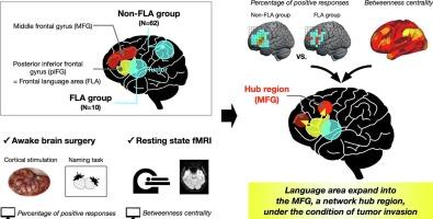
额叶语言区;左侧额下后回(pIFG)或布洛卡区),对语言处理至关重要,可以在病变进展中重组。虽然对侧半球的重组是众所周知的,但同侧半球的重组是如何发生的,特别是在病灶周围区域,尚不清楚。清醒手术期间的直接电刺激(DES)能够以高空间分辨率识别大脑区域与语言功能之间的因果关系。在这项研究中,我们研究了FLA同侧半球的皮质重组。本文对72例左脑胶质瘤患者进行了研究。根据病变是否包含pIFG将患者分为FLA组和非FLA组(n = 10和n = 62)。根据清醒手术指南的建议,所有患者在图片命名任务期间都进行了DES。一个子集在手术前接受静息状态功能MRI (rsfMRI)来计算中间性中心性,这是脑区域网络重要性的指标。DES显示,两组的pIFG对图片命名任务都表现出积极(受损)的反应。值得注意的是,与非FLA组相比,FLA组中额叶回(MFG)阳性反应的频率明显更高。基于rsfmri的网络分析显示,两组MFG的两个区域,一个在前部,另一个在后部,都比周围额叶区域表现出更高的中心性,尤其是后部。这些结果表明,在肿瘤进展后,语言区可以在病灶周围的MFG区域观察到,并提出了网络枢纽有助于维持脑病变后认知功能的可能性。
本文章由计算机程序翻译,如有差异,请以英文原文为准。

Broader functionality of language areas at the left middle frontal gyrus in patients with Broca’s area tumors

Broader functionality of language areas at the left middle frontal gyrus in patients with Broca’s area tumors
The frontal language area (FLA; left posterior inferior frontal gyrus [pIFG] or Broca’s area), critical for language processing can reorganize in response to lesion progression. While reorganization in the contralateral hemisphere is well known, how reorganization occurs within the ipsilateral hemisphere, especially in the perilesional region, remains unclear. Direct electrical stimulation (DES) during awake surgery enables identification of causal relationships between brain regions and language functions with high spatial resolution. In this study, we investigated cortical reorganization within the ipsilateral hemisphere of the FLA. Seventy-two patients with left hemisphere gliomas were studied. Patients were divided into FLA and non-FLA groups based on whether lesions included the pIFG (n = 10 and n = 62, respectively). All patients underwent DES during a picture-naming task, as recommended by awake surgery guidelines. A subset also underwent resting-state functional MRI (rsfMRI) before surgery to calculate betweenness centrality, an index of network importance of brain areas. DES revealed that the pIFG exhibited positive (impaired) responses to the picture-naming task in both groups. Notably, the frequency of positive responses in the middle frontal gyrus (MFG) was significantly higher in the FLA group than in the non-FLA group. RsfMRI-based network analyses revealed that two areas in the MFG, one in the anterior part and the other in the posterior part, showed higher centrality than surrounding frontal areas in both groups, especially the posterior one. These results suggest that language areas can be observed in the perilesional MFG regions following tumor progression, and raise the possibility that network hubs contribute to maintaining cognitive functions after brain lesions.
求助全文
通过发布文献求助,成功后即可免费获取论文全文。 去求助
来源期刊
Neuroimage-Clinical
Neuroimage-Clinical NEUROIMAGING-
CiteScore
7.50
自引率
4.80%
发文量
368
审稿时长
52 days
期刊介绍: NeuroImage: Clinical, a journal of diseases, disorders and syndromes involving the Nervous System, provides a vehicle for communicating important advances in the study of abnormal structure-function relationships of the human nervous system based on imaging. The focus of NeuroImage: Clinical is on defining changes to the brain associated with primary neurologic and psychiatric diseases and disorders of the nervous system as well as behavioral syndromes and developmental conditions. The main criterion for judging papers is the extent of scientific advancement in the understanding of the pathophysiologic mechanisms of diseases and disorders, in identification of functional models that link clinical signs and symptoms with brain function and in the creation of image based tools applicable to a broad range of clinical needs including diagnosis, monitoring and tracking of illness, predicting therapeutic response and development of new treatments. Papers dealing with structure and function in animal models will also be considered if they reveal mechanisms that can be readily translated to human conditions.
×
引用
GB/T 7714-2015
复制
MLA
复制
APA
复制
导出至
BibTeX EndNote RefMan NoteFirst NoteExpress
×
提示
您的信息不完整,为了账户安全,请先补充。
现在去补充
×
提示
您因"违规操作"
具体请查看互助需知
我知道了
×
提示
确定
请完成安全验证×
copy
已复制链接
快去分享给好友吧!
我知道了
右上角分享
点击右上角分享
0
联系我们:info@booksci.cn Book学术提供免费学术资源搜索服务,方便国内外学者检索中英文文献。致力于提供最便捷和优质的服务体验。 Copyright © 2023 布克学术 All rights reserved.
京ICP备2023020795号-1
ghs 京公网安备 11010802042870号
Book学术文献互助
Book学术文献互助群
群 号:604180095
Book学术官方微信