Ling Wang , Beining Yang , Qunya Qi , Haotian Xin , Yu Wang , Yulong Jia , Qian Chen , Weimin Zheng , Xin Chen , Tengfei Liang , Chuchu Sun , Jubao Du , Baowei Li , Jie Lu , Nan Chen
{"title":"小儿脊髓损伤患者脑功能重组及运动想象训练对其影响的初步研究","authors":"Ling Wang , Beining Yang , Qunya Qi , Haotian Xin , Yu Wang , Yulong Jia , Qian Chen , Weimin Zheng , Xin Chen , Tengfei Liang , Chuchu Sun , Jubao Du , Baowei Li , Jie Lu , Nan Chen","doi":"10.1016/j.ibneur.2025.08.003","DOIUrl":null,"url":null,"abstract":"<div><div>To study the brain functional alterations of children after spinal cord injury (SCI) and explore their changes after motor imagery training (MIT), revealing brain functional reorganizations in pediatric SCI and finding possible neural mechanisms of MIT. Thirty pediatric SCI patients and 30 age- and gender-matched healthy controls (HCs) were recruited. Brain resting-state functional MRI images of all subjects were obtained using a 3.0 Tesla MRI system. Subsequently, eight of the patients completed a 4-week MIT, and then functional MRI scans were conducted once again. Then two-sample t-tests were used to compare amplitude of low frequency (ALFF), fractional ALFF (fALFF), regional homogeneity (ReHo) between groups at baseline, and paired t-tests were used to investigate the changes in ALFF, fALFF and ReHo of patients before and after the treatment. Compared with HCs, the patients showed decreased ALFF and/or ReHo in bilateral postcentral gyrus (S1) and right orbitofrontal cortex, while increased ALFF and/or ReHo in the bilateral cerebellar lobules IV-VI, thalamus, left middle cingulate cortex (MCC), cerebellar Crus II, and right parahippocampal gyrus, caudate nucleus, middle temporal gyrus (MTG). Compared with those before MIT, the patients showed significantly increased ALFF in the right S1 after the treatment. These findings demonstrated brain functional reorganization in sensoriomotor, cognitive-emotional and auditory/language related regions, and MIT may promote the rehabilitation by reversing the functionally reorganized sensoriomotor areas, which may provide a possible mechanism for MIT.</div></div>","PeriodicalId":13195,"journal":{"name":"IBRO Neuroscience Reports","volume":"19 ","pages":"Pages 391-399"},"PeriodicalIF":2.9000,"publicationDate":"2025-08-07","publicationTypes":"Journal Article","fieldsOfStudy":null,"isOpenAccess":false,"openAccessPdf":"","citationCount":"0","resultStr":"{\"title\":\"Brain functional reorganization in pediatric patients with spinal cord injury and the impact of motor imagery training on it: a preliminary study\",\"authors\":\"Ling Wang , Beining Yang , Qunya Qi , Haotian Xin , Yu Wang , Yulong Jia , Qian Chen , Weimin Zheng , Xin Chen , Tengfei Liang , Chuchu Sun , Jubao Du , Baowei Li , Jie Lu , Nan Chen\",\"doi\":\"10.1016/j.ibneur.2025.08.003\",\"DOIUrl\":null,\"url\":null,\"abstract\":\"<div><div>To study the brain functional alterations of children after spinal cord injury (SCI) and explore their changes after motor imagery training (MIT), revealing brain functional reorganizations in pediatric SCI and finding possible neural mechanisms of MIT. Thirty pediatric SCI patients and 30 age- and gender-matched healthy controls (HCs) were recruited. Brain resting-state functional MRI images of all subjects were obtained using a 3.0 Tesla MRI system. Subsequently, eight of the patients completed a 4-week MIT, and then functional MRI scans were conducted once again. Then two-sample t-tests were used to compare amplitude of low frequency (ALFF), fractional ALFF (fALFF), regional homogeneity (ReHo) between groups at baseline, and paired t-tests were used to investigate the changes in ALFF, fALFF and ReHo of patients before and after the treatment. Compared with HCs, the patients showed decreased ALFF and/or ReHo in bilateral postcentral gyrus (S1) and right orbitofrontal cortex, while increased ALFF and/or ReHo in the bilateral cerebellar lobules IV-VI, thalamus, left middle cingulate cortex (MCC), cerebellar Crus II, and right parahippocampal gyrus, caudate nucleus, middle temporal gyrus (MTG). Compared with those before MIT, the patients showed significantly increased ALFF in the right S1 after the treatment. These findings demonstrated brain functional reorganization in sensoriomotor, cognitive-emotional and auditory/language related regions, and MIT may promote the rehabilitation by reversing the functionally reorganized sensoriomotor areas, which may provide a possible mechanism for MIT.</div></div>\",\"PeriodicalId\":13195,\"journal\":{\"name\":\"IBRO Neuroscience Reports\",\"volume\":\"19 \",\"pages\":\"Pages 391-399\"},\"PeriodicalIF\":2.9000,\"publicationDate\":\"2025-08-07\",\"publicationTypes\":\"Journal Article\",\"fieldsOfStudy\":null,\"isOpenAccess\":false,\"openAccessPdf\":\"\",\"citationCount\":\"0\",\"resultStr\":null,\"platform\":\"Semanticscholar\",\"paperid\":null,\"PeriodicalName\":\"IBRO Neuroscience Reports\",\"FirstCategoryId\":\"1085\",\"ListUrlMain\":\"https://www.sciencedirect.com/science/article/pii/S2667242125001198\",\"RegionNum\":0,\"RegionCategory\":null,\"ArticlePicture\":[],\"TitleCN\":null,\"AbstractTextCN\":null,\"PMCID\":null,\"EPubDate\":\"\",\"PubModel\":\"\",\"JCR\":\"Q3\",\"JCRName\":\"NEUROSCIENCES\",\"Score\":null,\"Total\":0}","platform":"Semanticscholar","paperid":null,"PeriodicalName":"IBRO Neuroscience Reports","FirstCategoryId":"1085","ListUrlMain":"https://www.sciencedirect.com/science/article/pii/S2667242125001198","RegionNum":0,"RegionCategory":null,"ArticlePicture":[],"TitleCN":null,"AbstractTextCN":null,"PMCID":null,"EPubDate":"","PubModel":"","JCR":"Q3","JCRName":"NEUROSCIENCES","Score":null,"Total":0}
引用次数: 0
摘要
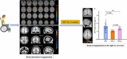
研究脊髓损伤(SCI)后儿童脑功能的变化,探讨运动意象训练(MIT)后儿童脑功能的变化,揭示小儿脊髓损伤后脑功能的重组,寻找可能的MIT神经机制。招募了30名小儿脊髓损伤患者和30名年龄和性别匹配的健康对照(hc)。采用3.0 Tesla MRI系统获得所有受试者的脑静息状态功能MRI图像。随后,8名患者完成了为期4周的MIT,然后再次进行功能性MRI扫描。然后采用双样本t检验比较各组基线时低频振幅(ALFF)、分数ALFF (fALFF)、区域均匀性(ReHo),采用配对t检验比较治疗前后患者ALFF、fALFF、ReHo的变化。与hc相比,患者双侧中央后回(S1)和右侧眶额皮质的ALFF和/或ReHo降低,而双侧小脑IV-VI小叶、丘脑、左侧中扣带皮层(MCC)、小脑小腿II、右侧海马旁回、尾状核、颞中回(MTG)的ALFF和/或ReHo升高。与治疗前相比,治疗后患者右侧S1区ALFF明显升高。这些结果表明,在感觉运动区、认知-情绪区和听觉/语言相关区域存在脑功能重组,而MIT可能通过逆转感觉运动区功能重组来促进康复,这可能为MIT的机制提供了可能。
Brain functional reorganization in pediatric patients with spinal cord injury and the impact of motor imagery training on it: a preliminary study
To study the brain functional alterations of children after spinal cord injury (SCI) and explore their changes after motor imagery training (MIT), revealing brain functional reorganizations in pediatric SCI and finding possible neural mechanisms of MIT. Thirty pediatric SCI patients and 30 age- and gender-matched healthy controls (HCs) were recruited. Brain resting-state functional MRI images of all subjects were obtained using a 3.0 Tesla MRI system. Subsequently, eight of the patients completed a 4-week MIT, and then functional MRI scans were conducted once again. Then two-sample t-tests were used to compare amplitude of low frequency (ALFF), fractional ALFF (fALFF), regional homogeneity (ReHo) between groups at baseline, and paired t-tests were used to investigate the changes in ALFF, fALFF and ReHo of patients before and after the treatment. Compared with HCs, the patients showed decreased ALFF and/or ReHo in bilateral postcentral gyrus (S1) and right orbitofrontal cortex, while increased ALFF and/or ReHo in the bilateral cerebellar lobules IV-VI, thalamus, left middle cingulate cortex (MCC), cerebellar Crus II, and right parahippocampal gyrus, caudate nucleus, middle temporal gyrus (MTG). Compared with those before MIT, the patients showed significantly increased ALFF in the right S1 after the treatment. These findings demonstrated brain functional reorganization in sensoriomotor, cognitive-emotional and auditory/language related regions, and MIT may promote the rehabilitation by reversing the functionally reorganized sensoriomotor areas, which may provide a possible mechanism for MIT.