评估烟雾病的t MRA技术:4D-ASL-MRA与3D-TOF-MRA。
IF 3.3
3区 医学
Q2 CLINICAL NEUROLOGY
引用次数: 0
摘要
目的:5.0T时三维飞行时间磁共振血管造影(3D-TOF-MRA)显示出与7.0T相当的小血管可视化能力,但尚未与5.0T时基于动脉自旋标记(ASL)的四维磁共振血管造影(4D-ASL-MRA)进行比较。本研究旨在比较这两种MRA技术在5.0T下评估烟雾病(MMD)的性能。方法:本回顾性研究包括20例连续接受6次4D-ASL-MRA (100ms至1800ms)和5.0T 3D-TOF-MRA的烟雾病患者。分析包括铃木分级、噪声对比比(CNR)、大脑中动脉(MCA)分支数量、颈动脉终末(ICA)、ICA终末周围狭窄、MCA远端、烟雾血管和小脑膜吻合(LMA)侧支血管的图像评分。结果:分析了20例患者(女性10例,33±8岁)30个大脑半球。与3D-TOF-MRA相比,标记时间为900ms至1800ms的4D-ASL-MRA在ICA末端、ICA周围狭窄和LMA的可视化评分上优于3D-TOF-MRA。4.00±0.00 vs 3.50±0.68,3.00±0.00 vs 2.68±0.55,2.37±1.19 vs 1.40±0.97,分别,p < 0.05),在1800 ms显示更高的中国北车和4 d-asl-mra M4段(45.84±20.28 vs 27.54±24.46,p < 0.001),但低M1, M3段(65.61±36.22 vs 173.58±148.25,48.89±29.44 vs 122.86±104.23,44.68±30.05 vs 78.36±72.64,分别为p < 0.05),可见远MCA分支(24.83±5.49 vs 15.03±5.99,pConclusion:在5.0T时,4D-ASL-MRA显示出MMD末端ICA,远端MCA和侧支血管的优越可视化,并且比3D-TOF-MRA更准确地分期MMD。本文章由计算机程序翻译,如有差异,请以英文原文为准。
5.0T MRA techniques for evaluating Moyamoya disease: 4D-ASL-MRA vs. 3D-TOF-MRA
Purpose
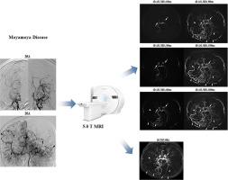
Three-dimensional time-of-flight MR-angiography (3D-TOF-MRA) at 5.0T showed comparable capacity to 7.0T for visualizing small vessels, but has not yet been compared with arterial spin labeling (ASL)-based four-dimensional MR-angiography (4D-ASL-MRA) at 5.0T. This study aimed to compare the performance of these two MRA techniques at 5.0T in evaluating Moyamoya disease (MMD).
Methods
This retrospective study included 20 consecutive MMD patients who underwent 4D-ASL-MRA with six labeling times (100 ms to 1800 ms) and 3D-TOF-MRA at 5.0T Analyses included Suzuki grades, contrast-to-noise ratio (CNR), number of branches in middle cerebral artery (MCA), and image scores of terminal internal carotid arteries (ICA), stenosis around terminal ICA, distal MCA, Moyamoya vessels, and leptomeningeal anastomosis (LMA) collateral vessels.
Results
Twenty patients (10 females, 33 ± 8 years) with 30 cerebral hemispheres were analyzed. Compared to 3D-TOF-MRA, 4D-ASL-MRA with 900 ms to 1800 ms labeling times demonstrated superior visualization scores for terminal ICA, stenosis around terminal ICA, and LMA (4D-ASL-MRA with 900 ms vs 3D-TOF-MRA: 4.00 ± 0.00 vs 3.50 ± 0.68, 3.00 ± 0.00 vs 2.68 ± 0.55, 2.37 ± 1.19 vs 1.40 ± 0.97, respectively, all p < 0.05), and 4D-ASL-MRA at 1800 ms showed higher CNR in the M4 segment (45.84 ± 20.28 vs 27.54 ± 24.46, p < 0.001) but lower in M1 to M3 segments (65.61 ± 36.22 vs 173.58 ± 148.25, 48.89 ± 29.44 vs 122.86 ± 104.23, 44.68 ± 30.05 vs 78.36 ± 72.64, respectively, all p < 0.05) and more visible distal MCA branches (24.83 ± 5.49 vs 15.03 ± 5.99, p < 0.001). The inter-modality agreement on Suzuki grades between 4D-ASL-MRA and digital subtraction angiography (DSA) was excellent (κ= 0.88), outperforming that between 3D-TOF-MRA and DSA (κ= 0.57).
Conclusion
At 5.0T, 4D-ASL-MRA demonstrated superior visualization of terminal ICA, distal MCA, and collateral vessels in MMD, as well as staging MMD more accurately than 3D-TOF-MRA.
求助全文
通过发布文献求助,成功后即可免费获取论文全文。
去求助
来源期刊
Journal of Neuroradiology
医学-核医学
CiteScore
6.10
自引率
5.70%
发文量
142
审稿时长
6-12 weeks
期刊介绍:
The Journal of Neuroradiology is a peer-reviewed journal, publishing worldwide clinical and basic research in the field of diagnostic and Interventional neuroradiology, translational and molecular neuroimaging, and artificial intelligence in neuroradiology.
The Journal of Neuroradiology considers for publication articles, reviews, technical notes and letters to the editors (correspondence section), provided that the methodology and scientific content are of high quality, and that the results will have substantial clinical impact and/or physiological importance.
 求助内容:
求助内容: 应助结果提醒方式:


