{"title":"Klippel-Trenaunay综合征孤立性结肠受累:一例广泛血管畸形。","authors":"Nurbek Ilyassov, Yerzhan Shayakhmetov, Anuar Abdikarimov, Erlan Nurgaliev, Saken Saberbekov, Rakymzhan Aralbayev, Aiman Tokusheva, Vitaliy Kalina","doi":"10.2147/IMCRJ.S535985","DOIUrl":null,"url":null,"abstract":"<p><strong>Background: </strong>Rectal vascular malformations associated with Klippel-Trenaunay syndrome are exceedingly rare. While their diagnosis may be straightforward when characteristic features are present, such cases remain of significant educational value due to their unusual presentation and potential for misdiagnosis.</p><p><strong>Case presentation: </strong>Our study aimed to describe a clinical case of a 45-year old female patient. There were complaints of pain in the perianal area and periodic bleeding during/after defecation, prolapse of hemorrhoids, general weakness. Colonoscopy revealed varicose veins of the rectum, hemangioma of the rectal mucosa, and chronic internal hemorrhoids. Multislice computed tomography of the pelvic organs with intravenous bolus contrast was performed. The preliminary clinical diagnosis was hemangioma of the rectal mucosa, and vascular malformation of the rectum was considered operable. In our clinic, the patient underwent several-stage surgery: (1) implantation of a temporary vena cava filter into the inferior vena cava, (2) two weeks later laparoscopic-assisted anterior resection of the rectum with preventive transverse colostomy and demucosation of the rectal mucosa, (3) after 7 months following discharge, the closure of transverse colostomy. Pathological examination of the gross specimen revealed that mucous membrane of the colon in a section extending 12.0 cm up to the distal edge of the resection is compacted, coarsely lumpy, grayish-bluish in color, with multiple blood-filled cavities. The postoperative period proceeded smoothly. Oral nutrition and patient's activity began on the 1st day. Primary healing of postoperative wounds was occurred. The patient was discharged in satisfactory condition on the 6th day post-surgery.</p><p><strong>Conclusion: </strong>This clinical case is a case-of-interest due to its rare localization, asymptomatic course for a long time with a fairly large size of an excessively vascularized abnormal vascular formation. The appropriate approach to this pathology is laparoscopic surgery, which can be useful for both diagnostic and radical treatment of vascular malformations of the rectum.</p>","PeriodicalId":14337,"journal":{"name":"International Medical Case Reports Journal","volume":"18 ","pages":"997-1002"},"PeriodicalIF":0.7000,"publicationDate":"2025-08-07","publicationTypes":"Journal Article","fieldsOfStudy":null,"isOpenAccess":false,"openAccessPdf":"https://www.ncbi.nlm.nih.gov/pmc/articles/PMC12339189/pdf/","citationCount":"0","resultStr":"{\"title\":\"Isolated Colorectal Involvement in Klippel-Trenaunay Syndrome: A Case of Extensive Vascular Malformation.\",\"authors\":\"Nurbek Ilyassov, Yerzhan Shayakhmetov, Anuar Abdikarimov, Erlan Nurgaliev, Saken Saberbekov, Rakymzhan Aralbayev, Aiman Tokusheva, Vitaliy Kalina\",\"doi\":\"10.2147/IMCRJ.S535985\",\"DOIUrl\":null,\"url\":null,\"abstract\":\"<p><strong>Background: </strong>Rectal vascular malformations associated with Klippel-Trenaunay syndrome are exceedingly rare. While their diagnosis may be straightforward when characteristic features are present, such cases remain of significant educational value due to their unusual presentation and potential for misdiagnosis.</p><p><strong>Case presentation: </strong>Our study aimed to describe a clinical case of a 45-year old female patient. There were complaints of pain in the perianal area and periodic bleeding during/after defecation, prolapse of hemorrhoids, general weakness. Colonoscopy revealed varicose veins of the rectum, hemangioma of the rectal mucosa, and chronic internal hemorrhoids. Multislice computed tomography of the pelvic organs with intravenous bolus contrast was performed. The preliminary clinical diagnosis was hemangioma of the rectal mucosa, and vascular malformation of the rectum was considered operable. In our clinic, the patient underwent several-stage surgery: (1) implantation of a temporary vena cava filter into the inferior vena cava, (2) two weeks later laparoscopic-assisted anterior resection of the rectum with preventive transverse colostomy and demucosation of the rectal mucosa, (3) after 7 months following discharge, the closure of transverse colostomy. Pathological examination of the gross specimen revealed that mucous membrane of the colon in a section extending 12.0 cm up to the distal edge of the resection is compacted, coarsely lumpy, grayish-bluish in color, with multiple blood-filled cavities. The postoperative period proceeded smoothly. Oral nutrition and patient's activity began on the 1st day. Primary healing of postoperative wounds was occurred. The patient was discharged in satisfactory condition on the 6th day post-surgery.</p><p><strong>Conclusion: </strong>This clinical case is a case-of-interest due to its rare localization, asymptomatic course for a long time with a fairly large size of an excessively vascularized abnormal vascular formation. The appropriate approach to this pathology is laparoscopic surgery, which can be useful for both diagnostic and radical treatment of vascular malformations of the rectum.</p>\",\"PeriodicalId\":14337,\"journal\":{\"name\":\"International Medical Case Reports Journal\",\"volume\":\"18 \",\"pages\":\"997-1002\"},\"PeriodicalIF\":0.7000,\"publicationDate\":\"2025-08-07\",\"publicationTypes\":\"Journal Article\",\"fieldsOfStudy\":null,\"isOpenAccess\":false,\"openAccessPdf\":\"https://www.ncbi.nlm.nih.gov/pmc/articles/PMC12339189/pdf/\",\"citationCount\":\"0\",\"resultStr\":null,\"platform\":\"Semanticscholar\",\"paperid\":null,\"PeriodicalName\":\"International Medical Case Reports Journal\",\"FirstCategoryId\":\"1085\",\"ListUrlMain\":\"https://doi.org/10.2147/IMCRJ.S535985\",\"RegionNum\":0,\"RegionCategory\":null,\"ArticlePicture\":[],\"TitleCN\":null,\"AbstractTextCN\":null,\"PMCID\":null,\"EPubDate\":\"2025/1/1 0:00:00\",\"PubModel\":\"eCollection\",\"JCR\":\"Q3\",\"JCRName\":\"MEDICINE, GENERAL & INTERNAL\",\"Score\":null,\"Total\":0}","platform":"Semanticscholar","paperid":null,"PeriodicalName":"International Medical Case Reports Journal","FirstCategoryId":"1085","ListUrlMain":"https://doi.org/10.2147/IMCRJ.S535985","RegionNum":0,"RegionCategory":null,"ArticlePicture":[],"TitleCN":null,"AbstractTextCN":null,"PMCID":null,"EPubDate":"2025/1/1 0:00:00","PubModel":"eCollection","JCR":"Q3","JCRName":"MEDICINE, GENERAL & INTERNAL","Score":null,"Total":0}
Isolated Colorectal Involvement in Klippel-Trenaunay Syndrome: A Case of Extensive Vascular Malformation.
Background: Rectal vascular malformations associated with Klippel-Trenaunay syndrome are exceedingly rare. While their diagnosis may be straightforward when characteristic features are present, such cases remain of significant educational value due to their unusual presentation and potential for misdiagnosis.
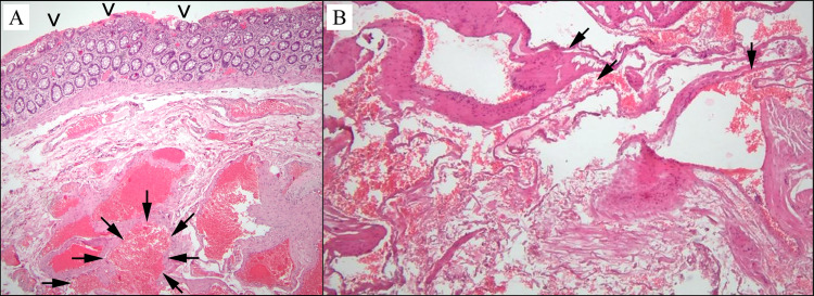
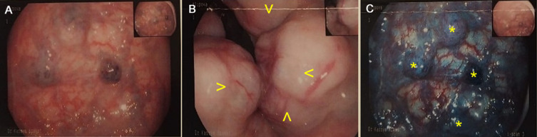
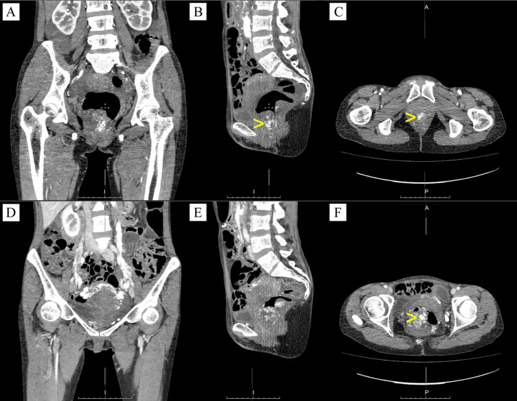
Case presentation: Our study aimed to describe a clinical case of a 45-year old female patient. There were complaints of pain in the perianal area and periodic bleeding during/after defecation, prolapse of hemorrhoids, general weakness. Colonoscopy revealed varicose veins of the rectum, hemangioma of the rectal mucosa, and chronic internal hemorrhoids. Multislice computed tomography of the pelvic organs with intravenous bolus contrast was performed. The preliminary clinical diagnosis was hemangioma of the rectal mucosa, and vascular malformation of the rectum was considered operable. In our clinic, the patient underwent several-stage surgery: (1) implantation of a temporary vena cava filter into the inferior vena cava, (2) two weeks later laparoscopic-assisted anterior resection of the rectum with preventive transverse colostomy and demucosation of the rectal mucosa, (3) after 7 months following discharge, the closure of transverse colostomy. Pathological examination of the gross specimen revealed that mucous membrane of the colon in a section extending 12.0 cm up to the distal edge of the resection is compacted, coarsely lumpy, grayish-bluish in color, with multiple blood-filled cavities. The postoperative period proceeded smoothly. Oral nutrition and patient's activity began on the 1st day. Primary healing of postoperative wounds was occurred. The patient was discharged in satisfactory condition on the 6th day post-surgery.
Conclusion: This clinical case is a case-of-interest due to its rare localization, asymptomatic course for a long time with a fairly large size of an excessively vascularized abnormal vascular formation. The appropriate approach to this pathology is laparoscopic surgery, which can be useful for both diagnostic and radical treatment of vascular malformations of the rectum.
期刊介绍:
International Medical Case Reports Journal is an international, peer-reviewed, open access, online journal publishing original case reports from all medical specialties. Submissions should not normally exceed 3,000 words or 4 published pages including figures, diagrams and references. As of 1st April 2019, the International Medical Case Reports Journal will no longer consider meta-analyses for publication.

 求助内容:
求助内容: 应助结果提醒方式:


