Irina A. Okkelman, Hang Zhou, Sergey M. Borisov, Angela C. Debruyne, Austin E. Y. T. Lefebvre, Marcelo Leomil Zoccoler, Linglong Chen, Bert Devriendt, Ruslan I. Dmitriev
{"title":"荧光寿命成像显微镜(FLIM)观察纳米塑料在活体肠道类器官中的内化和生物学影响","authors":"Irina A. Okkelman, Hang Zhou, Sergey M. Borisov, Angela C. Debruyne, Austin E. Y. T. Lefebvre, Marcelo Leomil Zoccoler, Linglong Chen, Bert Devriendt, Ruslan I. Dmitriev","doi":"10.1038/s41377-025-01949-0","DOIUrl":null,"url":null,"abstract":"<p>Increased micro- and nanoplastic (MNP) pollution poses significant health risks, yet the mechanisms of their accumulation and effects on absorptive tissues remain poorly understood. Addressing this knowledge gap requires tractable models coupled to dynamic live cell imaging methods, enabling multi-parameter single cell analysis. We report a new method combining adult stem cell-derived small intestinal organoid cultures with live fluorescence lifetime imaging microscopy (FLIM) to study MNP interactions with gut epithelium. To facilitate this, we optimized live imaging of porcine and mouse small intestinal organoids with an ‘apical-out’ topology. Subsequently, we produced a set of pristine MNPs based on PMMA and PS (<200 nm, doped with deep-red fluorescent dye) and evaluated their interaction with organoids displaying controlled epithelial polarity. We found that nanoparticles interacted differently with apical and basal membranes of the organoids and showed a species-specific pattern of cellular uptake. Using a phasor analysis approach, we demonstrate improved sensitivity of FLIM over conventional intensity-based microscopy. The resulting ‘fluorescence lifetime barcoding’ enabled distinguishing of different types of MNP and their interaction sites within organoids. Finally, we studied short (1 day)- and long (3 day)-term exposure effects of PMMA and PS-based MNPs on mitochondrial function, total cell energy budget and epithelial inflammation. We found that even pristine MNPs could disrupt chemokine production and mitochondrial membrane potential in intestinal epithelial cells. The presented FLIM approach will advance the study of MNP toxicity, their biological impacts on gastrointestinal tissue and enable the tracing of other fluorescent nanoparticles in live organoid and 3D ex vivo systems.</p>","PeriodicalId":18069,"journal":{"name":"Light-Science & Applications","volume":"42 1","pages":""},"PeriodicalIF":23.4000,"publicationDate":"2025-08-12","publicationTypes":"Journal Article","fieldsOfStudy":null,"isOpenAccess":false,"openAccessPdf":"","citationCount":"0","resultStr":"{\"title\":\"Visualizing the internalization and biological impact of nanoplastics in live intestinal organoids by Fluorescence Lifetime Imaging Microscopy (FLIM)\",\"authors\":\"Irina A. Okkelman, Hang Zhou, Sergey M. Borisov, Angela C. Debruyne, Austin E. Y. T. Lefebvre, Marcelo Leomil Zoccoler, Linglong Chen, Bert Devriendt, Ruslan I. Dmitriev\",\"doi\":\"10.1038/s41377-025-01949-0\",\"DOIUrl\":null,\"url\":null,\"abstract\":\"<p>Increased micro- and nanoplastic (MNP) pollution poses significant health risks, yet the mechanisms of their accumulation and effects on absorptive tissues remain poorly understood. Addressing this knowledge gap requires tractable models coupled to dynamic live cell imaging methods, enabling multi-parameter single cell analysis. We report a new method combining adult stem cell-derived small intestinal organoid cultures with live fluorescence lifetime imaging microscopy (FLIM) to study MNP interactions with gut epithelium. To facilitate this, we optimized live imaging of porcine and mouse small intestinal organoids with an ‘apical-out’ topology. Subsequently, we produced a set of pristine MNPs based on PMMA and PS (<200 nm, doped with deep-red fluorescent dye) and evaluated their interaction with organoids displaying controlled epithelial polarity. We found that nanoparticles interacted differently with apical and basal membranes of the organoids and showed a species-specific pattern of cellular uptake. Using a phasor analysis approach, we demonstrate improved sensitivity of FLIM over conventional intensity-based microscopy. The resulting ‘fluorescence lifetime barcoding’ enabled distinguishing of different types of MNP and their interaction sites within organoids. Finally, we studied short (1 day)- and long (3 day)-term exposure effects of PMMA and PS-based MNPs on mitochondrial function, total cell energy budget and epithelial inflammation. We found that even pristine MNPs could disrupt chemokine production and mitochondrial membrane potential in intestinal epithelial cells. The presented FLIM approach will advance the study of MNP toxicity, their biological impacts on gastrointestinal tissue and enable the tracing of other fluorescent nanoparticles in live organoid and 3D ex vivo systems.</p>\",\"PeriodicalId\":18069,\"journal\":{\"name\":\"Light-Science & Applications\",\"volume\":\"42 1\",\"pages\":\"\"},\"PeriodicalIF\":23.4000,\"publicationDate\":\"2025-08-12\",\"publicationTypes\":\"Journal Article\",\"fieldsOfStudy\":null,\"isOpenAccess\":false,\"openAccessPdf\":\"\",\"citationCount\":\"0\",\"resultStr\":null,\"platform\":\"Semanticscholar\",\"paperid\":null,\"PeriodicalName\":\"Light-Science & Applications\",\"FirstCategoryId\":\"1089\",\"ListUrlMain\":\"https://doi.org/10.1038/s41377-025-01949-0\",\"RegionNum\":0,\"RegionCategory\":null,\"ArticlePicture\":[],\"TitleCN\":null,\"AbstractTextCN\":null,\"PMCID\":null,\"EPubDate\":\"\",\"PubModel\":\"\",\"JCR\":\"Q1\",\"JCRName\":\"OPTICS\",\"Score\":null,\"Total\":0}","platform":"Semanticscholar","paperid":null,"PeriodicalName":"Light-Science & Applications","FirstCategoryId":"1089","ListUrlMain":"https://doi.org/10.1038/s41377-025-01949-0","RegionNum":0,"RegionCategory":null,"ArticlePicture":[],"TitleCN":null,"AbstractTextCN":null,"PMCID":null,"EPubDate":"","PubModel":"","JCR":"Q1","JCRName":"OPTICS","Score":null,"Total":0}
Visualizing the internalization and biological impact of nanoplastics in live intestinal organoids by Fluorescence Lifetime Imaging Microscopy (FLIM)
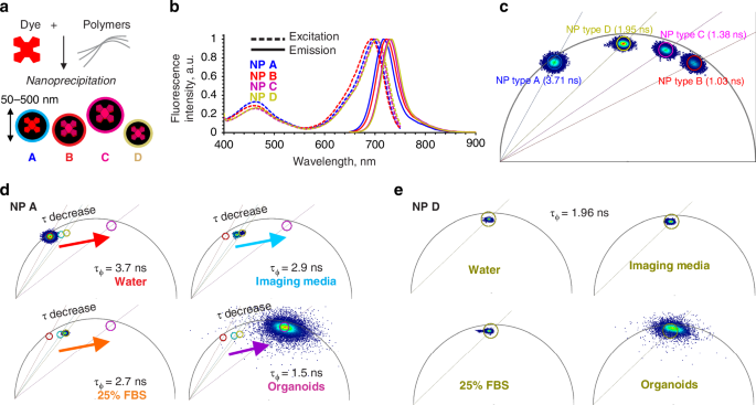
Increased micro- and nanoplastic (MNP) pollution poses significant health risks, yet the mechanisms of their accumulation and effects on absorptive tissues remain poorly understood. Addressing this knowledge gap requires tractable models coupled to dynamic live cell imaging methods, enabling multi-parameter single cell analysis. We report a new method combining adult stem cell-derived small intestinal organoid cultures with live fluorescence lifetime imaging microscopy (FLIM) to study MNP interactions with gut epithelium. To facilitate this, we optimized live imaging of porcine and mouse small intestinal organoids with an ‘apical-out’ topology. Subsequently, we produced a set of pristine MNPs based on PMMA and PS (<200 nm, doped with deep-red fluorescent dye) and evaluated their interaction with organoids displaying controlled epithelial polarity. We found that nanoparticles interacted differently with apical and basal membranes of the organoids and showed a species-specific pattern of cellular uptake. Using a phasor analysis approach, we demonstrate improved sensitivity of FLIM over conventional intensity-based microscopy. The resulting ‘fluorescence lifetime barcoding’ enabled distinguishing of different types of MNP and their interaction sites within organoids. Finally, we studied short (1 day)- and long (3 day)-term exposure effects of PMMA and PS-based MNPs on mitochondrial function, total cell energy budget and epithelial inflammation. We found that even pristine MNPs could disrupt chemokine production and mitochondrial membrane potential in intestinal epithelial cells. The presented FLIM approach will advance the study of MNP toxicity, their biological impacts on gastrointestinal tissue and enable the tracing of other fluorescent nanoparticles in live organoid and 3D ex vivo systems.
 求助内容:
求助内容: 应助结果提醒方式:


