{"title":"宫腔镜下继发性不孕症患者胎盘结节和斑块的临床意义:回顾性队列研究。","authors":"Rika Yamamichi, Mari Nomiyama, Kaoru Arima, Fumio Yamasaki, Kayoko Kojima, Michio Kitajima","doi":"10.4103/gmit.GMIT-D-24-00041","DOIUrl":null,"url":null,"abstract":"<p><strong>Objectives: </strong>To delineate the hysteroscopic findings of placental site nodule and plaque (PSNP) and determine its association with secondary infertility and chronic endometritis (CE).</p><p><strong>Materials and methods: </strong>We performed a retrospective cohort study of seven patients diagnosed with PSNP among women with secondary infertility mainly treated by assisted reproduction due to the variety of indications who underwent office mini-hysteroscopy with resection (MHR) followed by endometrial aspiration biopsy (EAB). Clinical backgrounds, specific hysteroscopic findings, and the sampling methods for the diagnosis of PSNP were analyzed. The presence of CE was diagnosed by CD138 immunostaining and relationship between PSNP and CE were evaluated. The clinical outcomes were recorded.</p><p><strong>Results: </strong>Multiple yellow-white colored protuberant lesion, micro polyps, and stalkless polyp were found and were resected under direct hysteroscopic observation. In five patients where PSNP was detected in both MHR and EAB samples, targeted biopsy using MHR within yellow-white colored protuberant lesion revealed PSNP. CD138 immunostaining showed marked plasma cell infiltration around the PSNP nodules in six patients and clinical CE were diagnosed in five patients. All women conceived spontaneously or with frozen-thaw embryo transfer after the procedures.</p><p><strong>Conclusion: </strong>Yellow-white colored protuberant lesion is likely to be specific hysteroscopic findings of PSNP. However, the possibility of PSNP covered by superficial endometrium or with nonspecific appearances should be considered. The presence of PSNP may interfere with fertility and that hysteroscopic detection and resection of these lesions may have clinical significance in women with secondary infertility. Inflammatory reaction caused by PSNP may provoke secondary CE in surrounding endometrium.</p>","PeriodicalId":45272,"journal":{"name":"Gynecology and Minimally Invasive Therapy-GMIT","volume":"14 3","pages":"207-214"},"PeriodicalIF":1.7000,"publicationDate":"2025-07-19","publicationTypes":"Journal Article","fieldsOfStudy":null,"isOpenAccess":false,"openAccessPdf":"https://www.ncbi.nlm.nih.gov/pmc/articles/PMC12334100/pdf/","citationCount":"0","resultStr":"{\"title\":\"The Clinical Significance of Placental Site Nodule and Plaque in Women with Secondary Infertility Treated with Office Hysteroscopic Surgery: The Case Series of Retrospective Cohort Study.\",\"authors\":\"Rika Yamamichi, Mari Nomiyama, Kaoru Arima, Fumio Yamasaki, Kayoko Kojima, Michio Kitajima\",\"doi\":\"10.4103/gmit.GMIT-D-24-00041\",\"DOIUrl\":null,\"url\":null,\"abstract\":\"<p><strong>Objectives: </strong>To delineate the hysteroscopic findings of placental site nodule and plaque (PSNP) and determine its association with secondary infertility and chronic endometritis (CE).</p><p><strong>Materials and methods: </strong>We performed a retrospective cohort study of seven patients diagnosed with PSNP among women with secondary infertility mainly treated by assisted reproduction due to the variety of indications who underwent office mini-hysteroscopy with resection (MHR) followed by endometrial aspiration biopsy (EAB). Clinical backgrounds, specific hysteroscopic findings, and the sampling methods for the diagnosis of PSNP were analyzed. The presence of CE was diagnosed by CD138 immunostaining and relationship between PSNP and CE were evaluated. The clinical outcomes were recorded.</p><p><strong>Results: </strong>Multiple yellow-white colored protuberant lesion, micro polyps, and stalkless polyp were found and were resected under direct hysteroscopic observation. In five patients where PSNP was detected in both MHR and EAB samples, targeted biopsy using MHR within yellow-white colored protuberant lesion revealed PSNP. CD138 immunostaining showed marked plasma cell infiltration around the PSNP nodules in six patients and clinical CE were diagnosed in five patients. All women conceived spontaneously or with frozen-thaw embryo transfer after the procedures.</p><p><strong>Conclusion: </strong>Yellow-white colored protuberant lesion is likely to be specific hysteroscopic findings of PSNP. However, the possibility of PSNP covered by superficial endometrium or with nonspecific appearances should be considered. The presence of PSNP may interfere with fertility and that hysteroscopic detection and resection of these lesions may have clinical significance in women with secondary infertility. Inflammatory reaction caused by PSNP may provoke secondary CE in surrounding endometrium.</p>\",\"PeriodicalId\":45272,\"journal\":{\"name\":\"Gynecology and Minimally Invasive Therapy-GMIT\",\"volume\":\"14 3\",\"pages\":\"207-214\"},\"PeriodicalIF\":1.7000,\"publicationDate\":\"2025-07-19\",\"publicationTypes\":\"Journal Article\",\"fieldsOfStudy\":null,\"isOpenAccess\":false,\"openAccessPdf\":\"https://www.ncbi.nlm.nih.gov/pmc/articles/PMC12334100/pdf/\",\"citationCount\":\"0\",\"resultStr\":null,\"platform\":\"Semanticscholar\",\"paperid\":null,\"PeriodicalName\":\"Gynecology and Minimally Invasive Therapy-GMIT\",\"FirstCategoryId\":\"1085\",\"ListUrlMain\":\"https://doi.org/10.4103/gmit.GMIT-D-24-00041\",\"RegionNum\":0,\"RegionCategory\":null,\"ArticlePicture\":[],\"TitleCN\":null,\"AbstractTextCN\":null,\"PMCID\":null,\"EPubDate\":\"2025/7/1 0:00:00\",\"PubModel\":\"eCollection\",\"JCR\":\"Q3\",\"JCRName\":\"OBSTETRICS & GYNECOLOGY\",\"Score\":null,\"Total\":0}","platform":"Semanticscholar","paperid":null,"PeriodicalName":"Gynecology and Minimally Invasive Therapy-GMIT","FirstCategoryId":"1085","ListUrlMain":"https://doi.org/10.4103/gmit.GMIT-D-24-00041","RegionNum":0,"RegionCategory":null,"ArticlePicture":[],"TitleCN":null,"AbstractTextCN":null,"PMCID":null,"EPubDate":"2025/7/1 0:00:00","PubModel":"eCollection","JCR":"Q3","JCRName":"OBSTETRICS & GYNECOLOGY","Score":null,"Total":0}
The Clinical Significance of Placental Site Nodule and Plaque in Women with Secondary Infertility Treated with Office Hysteroscopic Surgery: The Case Series of Retrospective Cohort Study.
Objectives: To delineate the hysteroscopic findings of placental site nodule and plaque (PSNP) and determine its association with secondary infertility and chronic endometritis (CE).
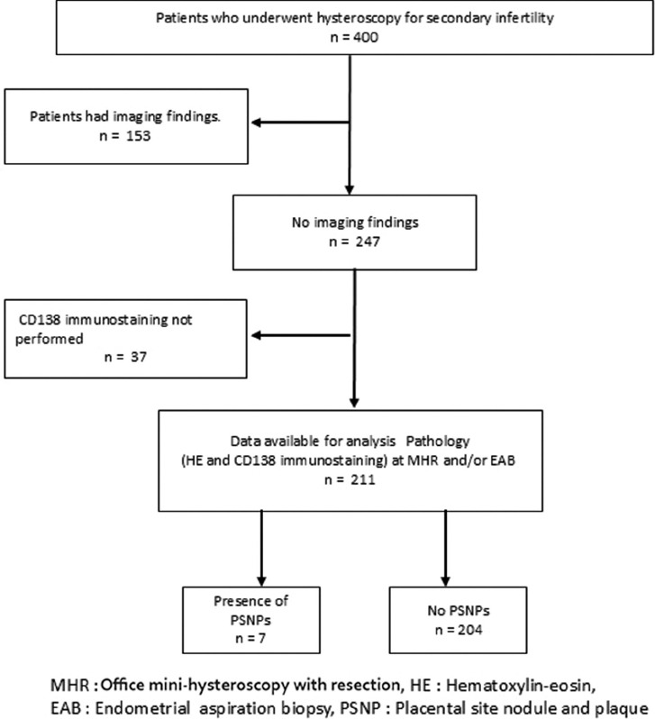
Materials and methods: We performed a retrospective cohort study of seven patients diagnosed with PSNP among women with secondary infertility mainly treated by assisted reproduction due to the variety of indications who underwent office mini-hysteroscopy with resection (MHR) followed by endometrial aspiration biopsy (EAB). Clinical backgrounds, specific hysteroscopic findings, and the sampling methods for the diagnosis of PSNP were analyzed. The presence of CE was diagnosed by CD138 immunostaining and relationship between PSNP and CE were evaluated. The clinical outcomes were recorded.
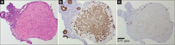
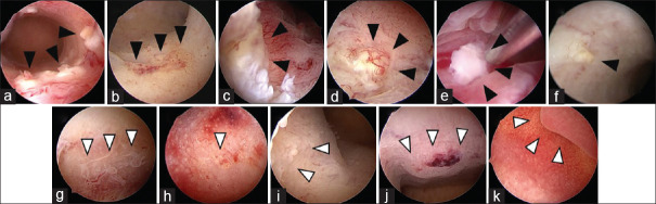
Results: Multiple yellow-white colored protuberant lesion, micro polyps, and stalkless polyp were found and were resected under direct hysteroscopic observation. In five patients where PSNP was detected in both MHR and EAB samples, targeted biopsy using MHR within yellow-white colored protuberant lesion revealed PSNP. CD138 immunostaining showed marked plasma cell infiltration around the PSNP nodules in six patients and clinical CE were diagnosed in five patients. All women conceived spontaneously or with frozen-thaw embryo transfer after the procedures.
Conclusion: Yellow-white colored protuberant lesion is likely to be specific hysteroscopic findings of PSNP. However, the possibility of PSNP covered by superficial endometrium or with nonspecific appearances should be considered. The presence of PSNP may interfere with fertility and that hysteroscopic detection and resection of these lesions may have clinical significance in women with secondary infertility. Inflammatory reaction caused by PSNP may provoke secondary CE in surrounding endometrium.

 求助内容:
求助内容: 应助结果提醒方式:


