Yao Xu, Yingjie Chen, Jiali Fan, Sasa Kou, Xinyu Zhuang, Bingyuan Zhou, Xiaofeng Zhang
{"title":"中国大陆法布里病的眼部和共焦表现:一项横断面对照研究。","authors":"Yao Xu, Yingjie Chen, Jiali Fan, Sasa Kou, Xinyu Zhuang, Bingyuan Zhou, Xiaofeng Zhang","doi":"10.1186/s13023-025-03940-9","DOIUrl":null,"url":null,"abstract":"<p><strong>Background: </strong>This cross-sectional controlled study aims to characterize ocular manifestations and corneal microstructure via in vivo confocal microscopy (IVCM) in mainland Chinese patients with Fabry disease (FD). We evaluated 30 FD patients (mean age: 38 ± 14.41 years; range: 10-60 years), divided equally into enzyme replacement therapy (ERT)-treated and untreated groups, alongside 30 age- and gender-matched healthy controls. Slit-lamp examinations assessed ocular manifestations, while IVCM was employed to analyze corneal microstructure.</p><p><strong>Results: </strong>Eighteen FD patients presented with corneal verticillata (CV) opacities. High-reflective intracellular inclusions were identified in the corneal basal epithelial cells in the majority of FD patients (22 out of 30). IVCM detected increased dendritic cells (DCs) in three FD patients. The nerve fiber layer showed an increased corneal nerve tortuosity coefficient (P < 0.001), decreased nerve fiber density (NFD) (P = 0.033), decreased nerve fiber length (NFL) (P = 0.012), and reduced fractal dimension (P = 0.010) in FD patients compared to healthy controls. Reduced transparency of the anterior corneal stroma and the presence of visible microdots were observed in 11 out of 30 FD patients. Endothelial morphological parameters in FD patients showed no obvious differences compared to healthy controls. α-galactosidase A (α-Gal A) activity was negatively correlated with Mainz Severity Score Index (MSSI) scores (P = 0.001), whereas plasma globotriaosylsphingosine (lyso-Gb3) levels and posterior capsular opacification exhibited a direct correlation with MSSI scores(P = 0.002). None of these changes showed significant differences in FD patients, regardless of ERT.</p><p><strong>Conclusions: </strong>This study substantially enhances our understanding of FD-associated ocular alterations in the mainland Chinese demographic. The presence of CV opacities, posterior capsular opacification, or distinct changes observed in IVCM offers the potential for early detection of FD. Additionally, there is a notable increase in DCs and a positive correlation between posterior capsular opacification and MSSI scores. These findings support the integration of ocular biomarker screening into standardized FD diagnostic protocols to facilitate pre-symptomatic interventions, particularly in familial risk cohorts.</p>","PeriodicalId":19651,"journal":{"name":"Orphanet Journal of Rare Diseases","volume":"20 1","pages":"417"},"PeriodicalIF":3.5000,"publicationDate":"2025-08-10","publicationTypes":"Journal Article","fieldsOfStudy":null,"isOpenAccess":false,"openAccessPdf":"https://www.ncbi.nlm.nih.gov/pmc/articles/PMC12337376/pdf/","citationCount":"0","resultStr":"{\"title\":\"Ocular and confocal manifestations of Mainland Chinese with Fabry disease: a cross-sectional controlled study.\",\"authors\":\"Yao Xu, Yingjie Chen, Jiali Fan, Sasa Kou, Xinyu Zhuang, Bingyuan Zhou, Xiaofeng Zhang\",\"doi\":\"10.1186/s13023-025-03940-9\",\"DOIUrl\":null,\"url\":null,\"abstract\":\"<p><strong>Background: </strong>This cross-sectional controlled study aims to characterize ocular manifestations and corneal microstructure via in vivo confocal microscopy (IVCM) in mainland Chinese patients with Fabry disease (FD). We evaluated 30 FD patients (mean age: 38 ± 14.41 years; range: 10-60 years), divided equally into enzyme replacement therapy (ERT)-treated and untreated groups, alongside 30 age- and gender-matched healthy controls. Slit-lamp examinations assessed ocular manifestations, while IVCM was employed to analyze corneal microstructure.</p><p><strong>Results: </strong>Eighteen FD patients presented with corneal verticillata (CV) opacities. High-reflective intracellular inclusions were identified in the corneal basal epithelial cells in the majority of FD patients (22 out of 30). IVCM detected increased dendritic cells (DCs) in three FD patients. The nerve fiber layer showed an increased corneal nerve tortuosity coefficient (P < 0.001), decreased nerve fiber density (NFD) (P = 0.033), decreased nerve fiber length (NFL) (P = 0.012), and reduced fractal dimension (P = 0.010) in FD patients compared to healthy controls. Reduced transparency of the anterior corneal stroma and the presence of visible microdots were observed in 11 out of 30 FD patients. Endothelial morphological parameters in FD patients showed no obvious differences compared to healthy controls. α-galactosidase A (α-Gal A) activity was negatively correlated with Mainz Severity Score Index (MSSI) scores (P = 0.001), whereas plasma globotriaosylsphingosine (lyso-Gb3) levels and posterior capsular opacification exhibited a direct correlation with MSSI scores(P = 0.002). None of these changes showed significant differences in FD patients, regardless of ERT.</p><p><strong>Conclusions: </strong>This study substantially enhances our understanding of FD-associated ocular alterations in the mainland Chinese demographic. The presence of CV opacities, posterior capsular opacification, or distinct changes observed in IVCM offers the potential for early detection of FD. Additionally, there is a notable increase in DCs and a positive correlation between posterior capsular opacification and MSSI scores. These findings support the integration of ocular biomarker screening into standardized FD diagnostic protocols to facilitate pre-symptomatic interventions, particularly in familial risk cohorts.</p>\",\"PeriodicalId\":19651,\"journal\":{\"name\":\"Orphanet Journal of Rare Diseases\",\"volume\":\"20 1\",\"pages\":\"417\"},\"PeriodicalIF\":3.5000,\"publicationDate\":\"2025-08-10\",\"publicationTypes\":\"Journal Article\",\"fieldsOfStudy\":null,\"isOpenAccess\":false,\"openAccessPdf\":\"https://www.ncbi.nlm.nih.gov/pmc/articles/PMC12337376/pdf/\",\"citationCount\":\"0\",\"resultStr\":null,\"platform\":\"Semanticscholar\",\"paperid\":null,\"PeriodicalName\":\"Orphanet Journal of Rare Diseases\",\"FirstCategoryId\":\"3\",\"ListUrlMain\":\"https://doi.org/10.1186/s13023-025-03940-9\",\"RegionNum\":2,\"RegionCategory\":\"医学\",\"ArticlePicture\":[],\"TitleCN\":null,\"AbstractTextCN\":null,\"PMCID\":null,\"EPubDate\":\"\",\"PubModel\":\"\",\"JCR\":\"Q2\",\"JCRName\":\"GENETICS & HEREDITY\",\"Score\":null,\"Total\":0}","platform":"Semanticscholar","paperid":null,"PeriodicalName":"Orphanet Journal of Rare Diseases","FirstCategoryId":"3","ListUrlMain":"https://doi.org/10.1186/s13023-025-03940-9","RegionNum":2,"RegionCategory":"医学","ArticlePicture":[],"TitleCN":null,"AbstractTextCN":null,"PMCID":null,"EPubDate":"","PubModel":"","JCR":"Q2","JCRName":"GENETICS & HEREDITY","Score":null,"Total":0}
Ocular and confocal manifestations of Mainland Chinese with Fabry disease: a cross-sectional controlled study.
Background: This cross-sectional controlled study aims to characterize ocular manifestations and corneal microstructure via in vivo confocal microscopy (IVCM) in mainland Chinese patients with Fabry disease (FD). We evaluated 30 FD patients (mean age: 38 ± 14.41 years; range: 10-60 years), divided equally into enzyme replacement therapy (ERT)-treated and untreated groups, alongside 30 age- and gender-matched healthy controls. Slit-lamp examinations assessed ocular manifestations, while IVCM was employed to analyze corneal microstructure.
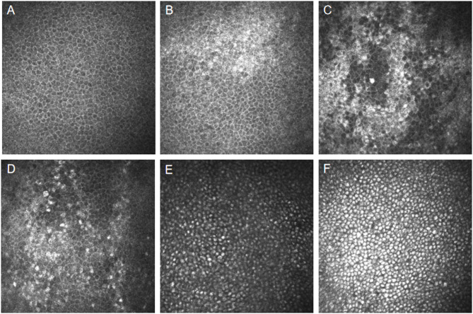
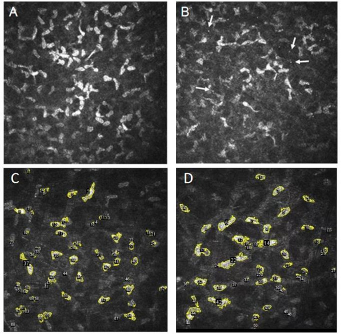
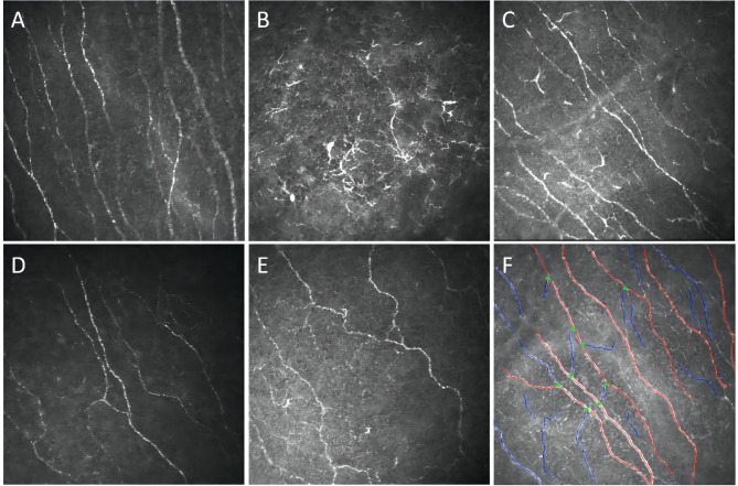
Results: Eighteen FD patients presented with corneal verticillata (CV) opacities. High-reflective intracellular inclusions were identified in the corneal basal epithelial cells in the majority of FD patients (22 out of 30). IVCM detected increased dendritic cells (DCs) in three FD patients. The nerve fiber layer showed an increased corneal nerve tortuosity coefficient (P < 0.001), decreased nerve fiber density (NFD) (P = 0.033), decreased nerve fiber length (NFL) (P = 0.012), and reduced fractal dimension (P = 0.010) in FD patients compared to healthy controls. Reduced transparency of the anterior corneal stroma and the presence of visible microdots were observed in 11 out of 30 FD patients. Endothelial morphological parameters in FD patients showed no obvious differences compared to healthy controls. α-galactosidase A (α-Gal A) activity was negatively correlated with Mainz Severity Score Index (MSSI) scores (P = 0.001), whereas plasma globotriaosylsphingosine (lyso-Gb3) levels and posterior capsular opacification exhibited a direct correlation with MSSI scores(P = 0.002). None of these changes showed significant differences in FD patients, regardless of ERT.
Conclusions: This study substantially enhances our understanding of FD-associated ocular alterations in the mainland Chinese demographic. The presence of CV opacities, posterior capsular opacification, or distinct changes observed in IVCM offers the potential for early detection of FD. Additionally, there is a notable increase in DCs and a positive correlation between posterior capsular opacification and MSSI scores. These findings support the integration of ocular biomarker screening into standardized FD diagnostic protocols to facilitate pre-symptomatic interventions, particularly in familial risk cohorts.
期刊介绍:
Orphanet Journal of Rare Diseases is an open access, peer-reviewed journal that encompasses all aspects of rare diseases and orphan drugs. The journal publishes high-quality reviews on specific rare diseases. In addition, the journal may consider articles on clinical trial outcome reports, either positive or negative, and articles on public health issues in the field of rare diseases and orphan drugs. The journal does not accept case reports.
 求助内容:
求助内容: 应助结果提醒方式:


