Leo Wan, Aileen Park, Lanah Almatroud, Amor Khachemoune
{"title":"皮肤纤维瘤:重新评估和更新综述。","authors":"Leo Wan, Aileen Park, Lanah Almatroud, Amor Khachemoune","doi":"10.2147/CCID.S526191","DOIUrl":null,"url":null,"abstract":"<p><p>Dermatofibromas (DF), also known as fibrous histiocytomas, are common benign cutaneous lesions characterized histologically by dermal proliferation of spindle-shaped fibrocytes, with the overlying epidermis often demonstrating hyperplasia with acanthosis, basal layer hyperpigmentation, and a characteristic \"collarette\" of epidermal hyperplasia surrounding the lesion. The etiology of DF remains debated, with theories ranging from reactive processes triggered by local trauma, such as insect bites, to spontaneous development. DF typically presents as a hyperkeratotic nodule or plaque, most often on the lower extremities, and can exhibit a wide spectrum of clinical appearances. Variants such as hemosiderotic, epithelioid, aneurysmal, and cellular DF show distinct clinical and histopathological features that may sometimes mimic malignant lesions. Dermoscopic findings can aid in diagnosis, although biopsy is often required for definitive classification. Discrepancies in the literature persist regarding the pathogenesis and classification of DF, and while DF is generally benign, rare cases of metastasizing DF have been reported. This review aims to provide an examination of DF, including its clinical manifestations, etiology, subtypes, histological features, and differential diagnoses. It also discusses dermoscopic findings, controversies in the literature, and current treatment options. A clearer understanding of its diverse presentations, along with refined diagnostic criteria, will enhance clinical management and treatment strategies.</p>","PeriodicalId":10447,"journal":{"name":"Clinical, Cosmetic and Investigational Dermatology","volume":"18 ","pages":"1873-1887"},"PeriodicalIF":2.2000,"publicationDate":"2025-08-04","publicationTypes":"Journal Article","fieldsOfStudy":null,"isOpenAccess":false,"openAccessPdf":"https://www.ncbi.nlm.nih.gov/pmc/articles/PMC12333637/pdf/","citationCount":"0","resultStr":"{\"title\":\"Dermatofibroma: Reappraisal and Updated Review.\",\"authors\":\"Leo Wan, Aileen Park, Lanah Almatroud, Amor Khachemoune\",\"doi\":\"10.2147/CCID.S526191\",\"DOIUrl\":null,\"url\":null,\"abstract\":\"<p><p>Dermatofibromas (DF), also known as fibrous histiocytomas, are common benign cutaneous lesions characterized histologically by dermal proliferation of spindle-shaped fibrocytes, with the overlying epidermis often demonstrating hyperplasia with acanthosis, basal layer hyperpigmentation, and a characteristic \\\"collarette\\\" of epidermal hyperplasia surrounding the lesion. The etiology of DF remains debated, with theories ranging from reactive processes triggered by local trauma, such as insect bites, to spontaneous development. DF typically presents as a hyperkeratotic nodule or plaque, most often on the lower extremities, and can exhibit a wide spectrum of clinical appearances. Variants such as hemosiderotic, epithelioid, aneurysmal, and cellular DF show distinct clinical and histopathological features that may sometimes mimic malignant lesions. Dermoscopic findings can aid in diagnosis, although biopsy is often required for definitive classification. Discrepancies in the literature persist regarding the pathogenesis and classification of DF, and while DF is generally benign, rare cases of metastasizing DF have been reported. This review aims to provide an examination of DF, including its clinical manifestations, etiology, subtypes, histological features, and differential diagnoses. It also discusses dermoscopic findings, controversies in the literature, and current treatment options. A clearer understanding of its diverse presentations, along with refined diagnostic criteria, will enhance clinical management and treatment strategies.</p>\",\"PeriodicalId\":10447,\"journal\":{\"name\":\"Clinical, Cosmetic and Investigational Dermatology\",\"volume\":\"18 \",\"pages\":\"1873-1887\"},\"PeriodicalIF\":2.2000,\"publicationDate\":\"2025-08-04\",\"publicationTypes\":\"Journal Article\",\"fieldsOfStudy\":null,\"isOpenAccess\":false,\"openAccessPdf\":\"https://www.ncbi.nlm.nih.gov/pmc/articles/PMC12333637/pdf/\",\"citationCount\":\"0\",\"resultStr\":null,\"platform\":\"Semanticscholar\",\"paperid\":null,\"PeriodicalName\":\"Clinical, Cosmetic and Investigational Dermatology\",\"FirstCategoryId\":\"3\",\"ListUrlMain\":\"https://doi.org/10.2147/CCID.S526191\",\"RegionNum\":4,\"RegionCategory\":\"医学\",\"ArticlePicture\":[],\"TitleCN\":null,\"AbstractTextCN\":null,\"PMCID\":null,\"EPubDate\":\"2025/1/1 0:00:00\",\"PubModel\":\"eCollection\",\"JCR\":\"Q3\",\"JCRName\":\"DERMATOLOGY\",\"Score\":null,\"Total\":0}","platform":"Semanticscholar","paperid":null,"PeriodicalName":"Clinical, Cosmetic and Investigational Dermatology","FirstCategoryId":"3","ListUrlMain":"https://doi.org/10.2147/CCID.S526191","RegionNum":4,"RegionCategory":"医学","ArticlePicture":[],"TitleCN":null,"AbstractTextCN":null,"PMCID":null,"EPubDate":"2025/1/1 0:00:00","PubModel":"eCollection","JCR":"Q3","JCRName":"DERMATOLOGY","Score":null,"Total":0}
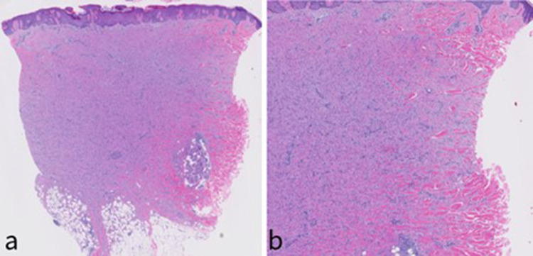
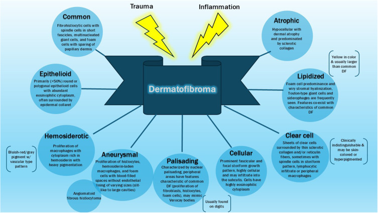
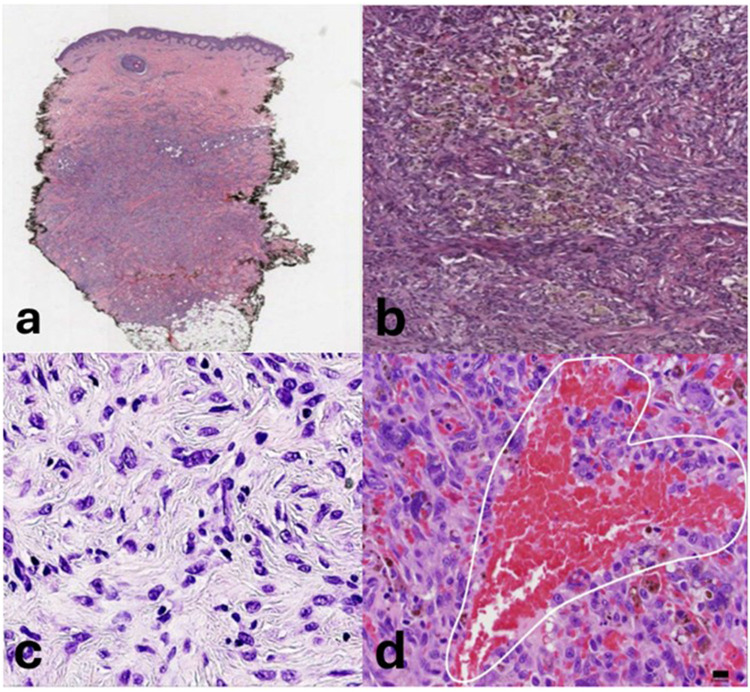
Dermatofibromas (DF), also known as fibrous histiocytomas, are common benign cutaneous lesions characterized histologically by dermal proliferation of spindle-shaped fibrocytes, with the overlying epidermis often demonstrating hyperplasia with acanthosis, basal layer hyperpigmentation, and a characteristic "collarette" of epidermal hyperplasia surrounding the lesion. The etiology of DF remains debated, with theories ranging from reactive processes triggered by local trauma, such as insect bites, to spontaneous development. DF typically presents as a hyperkeratotic nodule or plaque, most often on the lower extremities, and can exhibit a wide spectrum of clinical appearances. Variants such as hemosiderotic, epithelioid, aneurysmal, and cellular DF show distinct clinical and histopathological features that may sometimes mimic malignant lesions. Dermoscopic findings can aid in diagnosis, although biopsy is often required for definitive classification. Discrepancies in the literature persist regarding the pathogenesis and classification of DF, and while DF is generally benign, rare cases of metastasizing DF have been reported. This review aims to provide an examination of DF, including its clinical manifestations, etiology, subtypes, histological features, and differential diagnoses. It also discusses dermoscopic findings, controversies in the literature, and current treatment options. A clearer understanding of its diverse presentations, along with refined diagnostic criteria, will enhance clinical management and treatment strategies.
期刊介绍:
Clinical, Cosmetic and Investigational Dermatology is an international, peer-reviewed, open access journal that focuses on the latest clinical and experimental research in all aspects of skin disease and cosmetic interventions. Normal and pathological processes in skin development and aging, their modification and treatment, as well as basic research into histology of dermal and dermal structures that provide clinical insights and potential treatment options are key topics for the journal.
Patient satisfaction, preference, quality of life, compliance, persistence and their role in developing new management options to optimize outcomes for target conditions constitute major areas of interest.
The journal is characterized by the rapid reporting of clinical studies, reviews and original research in skin research and skin care.
All areas of dermatology will be covered; contributions will be welcomed from all clinicians and basic science researchers globally.
 求助内容:
求助内容: 应助结果提醒方式:


