{"title":"输尿管结石自发逆行迁移:罕见现象。","authors":"Tayyaba Kauser, Zain Ul Abdin, Wishah Urwatil Wusqa, Munna William, Momena Rashid, Rejina Chhetri","doi":"10.1097/MS9.0000000000003606","DOIUrl":null,"url":null,"abstract":"<p><strong>Introduction: </strong>While spontaneous anterograde passage of ureteric stones is well documented, retrograde migration-movement of the stone back towards the kidney is exceedingly rare. The mechanisms underlying this unusual migration pattern remain poorly understood, with only a few cases reported in the literature.</p><p><strong>Case presentation: </strong>A 23-year-old female presented with acute left flank pain, imaging for which revealed a calculus in the left proximal ureter. A repeat imaging after a few weeks of medical management revealed a calculus of the same dimension at the lower pole calyx of the left kidney, with no calculus present at the initial site, indicating retrograde migration of the calculus. The patient subsequently underwent extracorporeal shockwave lithotripsy with complete stone clearance.</p><p><strong>Discussion: </strong>Retrograde migration of ureteric calculi is a rare phenomenon. Proposed mechanisms include reverse ureteral peristalsis, proximal ureteral dilation, antiperistaltic waves due to irritant stimuli, and the effects of non-steroidal anti-inflammatory drugs (NSAIDs) on ureteric peristalsis. Awareness of this occurrence is important for appropriate diagnosis and management, as retrograde migration can affect treatment planning and outcomes.</p><p><strong>Conclusions: </strong>The case highlights an unusual presentation of urolithiasis that radiologists and urologists should be aware of as this can have significant implications for clinical management.</p>","PeriodicalId":8025,"journal":{"name":"Annals of Medicine and Surgery","volume":"87 8","pages":"5330-5332"},"PeriodicalIF":1.6000,"publicationDate":"2025-07-22","publicationTypes":"Journal Article","fieldsOfStudy":null,"isOpenAccess":false,"openAccessPdf":"https://www.ncbi.nlm.nih.gov/pmc/articles/PMC12333757/pdf/","citationCount":"0","resultStr":"{\"title\":\"Spontaneous retrograde migration of a ureteric calculus: a rare phenomenon.\",\"authors\":\"Tayyaba Kauser, Zain Ul Abdin, Wishah Urwatil Wusqa, Munna William, Momena Rashid, Rejina Chhetri\",\"doi\":\"10.1097/MS9.0000000000003606\",\"DOIUrl\":null,\"url\":null,\"abstract\":\"<p><strong>Introduction: </strong>While spontaneous anterograde passage of ureteric stones is well documented, retrograde migration-movement of the stone back towards the kidney is exceedingly rare. The mechanisms underlying this unusual migration pattern remain poorly understood, with only a few cases reported in the literature.</p><p><strong>Case presentation: </strong>A 23-year-old female presented with acute left flank pain, imaging for which revealed a calculus in the left proximal ureter. A repeat imaging after a few weeks of medical management revealed a calculus of the same dimension at the lower pole calyx of the left kidney, with no calculus present at the initial site, indicating retrograde migration of the calculus. The patient subsequently underwent extracorporeal shockwave lithotripsy with complete stone clearance.</p><p><strong>Discussion: </strong>Retrograde migration of ureteric calculi is a rare phenomenon. Proposed mechanisms include reverse ureteral peristalsis, proximal ureteral dilation, antiperistaltic waves due to irritant stimuli, and the effects of non-steroidal anti-inflammatory drugs (NSAIDs) on ureteric peristalsis. Awareness of this occurrence is important for appropriate diagnosis and management, as retrograde migration can affect treatment planning and outcomes.</p><p><strong>Conclusions: </strong>The case highlights an unusual presentation of urolithiasis that radiologists and urologists should be aware of as this can have significant implications for clinical management.</p>\",\"PeriodicalId\":8025,\"journal\":{\"name\":\"Annals of Medicine and Surgery\",\"volume\":\"87 8\",\"pages\":\"5330-5332\"},\"PeriodicalIF\":1.6000,\"publicationDate\":\"2025-07-22\",\"publicationTypes\":\"Journal Article\",\"fieldsOfStudy\":null,\"isOpenAccess\":false,\"openAccessPdf\":\"https://www.ncbi.nlm.nih.gov/pmc/articles/PMC12333757/pdf/\",\"citationCount\":\"0\",\"resultStr\":null,\"platform\":\"Semanticscholar\",\"paperid\":null,\"PeriodicalName\":\"Annals of Medicine and Surgery\",\"FirstCategoryId\":\"1085\",\"ListUrlMain\":\"https://doi.org/10.1097/MS9.0000000000003606\",\"RegionNum\":0,\"RegionCategory\":null,\"ArticlePicture\":[],\"TitleCN\":null,\"AbstractTextCN\":null,\"PMCID\":null,\"EPubDate\":\"2025/8/1 0:00:00\",\"PubModel\":\"eCollection\",\"JCR\":\"Q2\",\"JCRName\":\"MEDICINE, GENERAL & INTERNAL\",\"Score\":null,\"Total\":0}","platform":"Semanticscholar","paperid":null,"PeriodicalName":"Annals of Medicine and Surgery","FirstCategoryId":"1085","ListUrlMain":"https://doi.org/10.1097/MS9.0000000000003606","RegionNum":0,"RegionCategory":null,"ArticlePicture":[],"TitleCN":null,"AbstractTextCN":null,"PMCID":null,"EPubDate":"2025/8/1 0:00:00","PubModel":"eCollection","JCR":"Q2","JCRName":"MEDICINE, GENERAL & INTERNAL","Score":null,"Total":0}
Spontaneous retrograde migration of a ureteric calculus: a rare phenomenon.
Introduction: While spontaneous anterograde passage of ureteric stones is well documented, retrograde migration-movement of the stone back towards the kidney is exceedingly rare. The mechanisms underlying this unusual migration pattern remain poorly understood, with only a few cases reported in the literature.
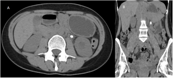
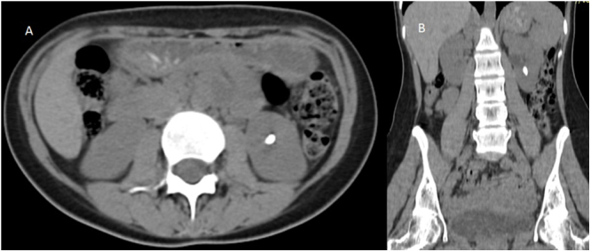
Case presentation: A 23-year-old female presented with acute left flank pain, imaging for which revealed a calculus in the left proximal ureter. A repeat imaging after a few weeks of medical management revealed a calculus of the same dimension at the lower pole calyx of the left kidney, with no calculus present at the initial site, indicating retrograde migration of the calculus. The patient subsequently underwent extracorporeal shockwave lithotripsy with complete stone clearance.
Discussion: Retrograde migration of ureteric calculi is a rare phenomenon. Proposed mechanisms include reverse ureteral peristalsis, proximal ureteral dilation, antiperistaltic waves due to irritant stimuli, and the effects of non-steroidal anti-inflammatory drugs (NSAIDs) on ureteric peristalsis. Awareness of this occurrence is important for appropriate diagnosis and management, as retrograde migration can affect treatment planning and outcomes.
Conclusions: The case highlights an unusual presentation of urolithiasis that radiologists and urologists should be aware of as this can have significant implications for clinical management.

 求助内容:
求助内容: 应助结果提醒方式:


