Amany F Elbehairy, Josephine H Naish, Hossein Baghertash, Geoff J M Parker, Christopher A Miller, Jørgen Vestbo, Alex R Horsley
{"title":"慢性阻塞性肺病患者的T2加权氧增强肺部MRI与静息和运动功能测量有关。","authors":"Amany F Elbehairy, Josephine H Naish, Hossein Baghertash, Geoff J M Parker, Christopher A Miller, Jørgen Vestbo, Alex R Horsley","doi":"10.1136/bmjresp-2024-002784","DOIUrl":null,"url":null,"abstract":"<p><strong>Background: </strong>T<sub>2</sub>*-weighted oxygen-enhanced MRI (T<sub>2</sub>*-OE-MRI) may directly assess pulmonary ventilation using oxygen as an inhaled tracer gas. It has shown promise in healthy volunteers (HVs) and cystic fibrosis but has yet to be demonstrated in patients with chronic obstructive pulmonary disease (COPD).</p><p><strong>Research question: </strong>To determine the feasibility and repeatability of T<sub>2</sub>*-OE-MRI in patients with COPD. To assess correlations between T<sub>2</sub>*-OE-MRI measurements of pulmonary ventilation, pulmonary function tests (PFTs) and measures of functional limitation.</p><p><strong>Study design and methods: </strong>13 patients with mild-to-severe COPD and 13 HVs underwent PFTs, lung clearance index (LCI) measurement, incremental exercise test (patients only) and two lung MRI scans at 3 T. For T<sub>2</sub>*-OE-MRI, participants were fitted with a non-rebreathing face mask and given 100% oxygen during image acquisition.</p><p><strong>Results: </strong>Patients (age: 63 (55-72) years, forced expiratory volume in 1 s (FEV<sub>1</sub>): 63 (36-79) %predicted, median (IQR)) had evidence of pulmonary gas trapping, small airway disease (SAD) and ventilation heterogeneity. During T<sub>2</sub>*-OE-MRI, the magnitude of the percentage difference between mean signal intensity at normoxia and hyperoxia (percent signal enhancement (PSE)) and the enhancing fraction (EF) were lower in patients versus HVs (2.77 (2.19-4.19) vs 5.34 (4.33-5.61) % and 0.74 (0.66-0.77) vs 0.89 (0.82-0.94), respectively, both p<0.001). Intraclass correlation coefficient values indicated moderate (0.74) and good (0.80) repeatability for PSE and EF, respectively. PSE and EF significantly correlated with FEV<sub>1</sub>, LCI and SAD indices, and in COPD, they correlated with measures of exercise capacity, dynamic hyperinflation and dyspnoea intensity during exercise.</p><p><strong>Interpretation: </strong>In patients with COPD, T<sub>2</sub>*-OE-MRI is feasible and repeatable and provides regional information on pulmonary ventilation that is linked with physiological measures of disease severity, functional limitation and exertional dyspnoea.</p>","PeriodicalId":9048,"journal":{"name":"BMJ Open Respiratory Research","volume":"12 1","pages":""},"PeriodicalIF":3.4000,"publicationDate":"2025-08-07","publicationTypes":"Journal Article","fieldsOfStudy":null,"isOpenAccess":false,"openAccessPdf":"https://www.ncbi.nlm.nih.gov/pmc/articles/PMC12336524/pdf/","citationCount":"0","resultStr":"{\"title\":\"T<sub>2</sub>*-weighted oxygen-enhanced pulmonary MRI in COPD is linked to resting and exertional functional measurements.\",\"authors\":\"Amany F Elbehairy, Josephine H Naish, Hossein Baghertash, Geoff J M Parker, Christopher A Miller, Jørgen Vestbo, Alex R Horsley\",\"doi\":\"10.1136/bmjresp-2024-002784\",\"DOIUrl\":null,\"url\":null,\"abstract\":\"<p><strong>Background: </strong>T<sub>2</sub>*-weighted oxygen-enhanced MRI (T<sub>2</sub>*-OE-MRI) may directly assess pulmonary ventilation using oxygen as an inhaled tracer gas. It has shown promise in healthy volunteers (HVs) and cystic fibrosis but has yet to be demonstrated in patients with chronic obstructive pulmonary disease (COPD).</p><p><strong>Research question: </strong>To determine the feasibility and repeatability of T<sub>2</sub>*-OE-MRI in patients with COPD. To assess correlations between T<sub>2</sub>*-OE-MRI measurements of pulmonary ventilation, pulmonary function tests (PFTs) and measures of functional limitation.</p><p><strong>Study design and methods: </strong>13 patients with mild-to-severe COPD and 13 HVs underwent PFTs, lung clearance index (LCI) measurement, incremental exercise test (patients only) and two lung MRI scans at 3 T. For T<sub>2</sub>*-OE-MRI, participants were fitted with a non-rebreathing face mask and given 100% oxygen during image acquisition.</p><p><strong>Results: </strong>Patients (age: 63 (55-72) years, forced expiratory volume in 1 s (FEV<sub>1</sub>): 63 (36-79) %predicted, median (IQR)) had evidence of pulmonary gas trapping, small airway disease (SAD) and ventilation heterogeneity. During T<sub>2</sub>*-OE-MRI, the magnitude of the percentage difference between mean signal intensity at normoxia and hyperoxia (percent signal enhancement (PSE)) and the enhancing fraction (EF) were lower in patients versus HVs (2.77 (2.19-4.19) vs 5.34 (4.33-5.61) % and 0.74 (0.66-0.77) vs 0.89 (0.82-0.94), respectively, both p<0.001). Intraclass correlation coefficient values indicated moderate (0.74) and good (0.80) repeatability for PSE and EF, respectively. PSE and EF significantly correlated with FEV<sub>1</sub>, LCI and SAD indices, and in COPD, they correlated with measures of exercise capacity, dynamic hyperinflation and dyspnoea intensity during exercise.</p><p><strong>Interpretation: </strong>In patients with COPD, T<sub>2</sub>*-OE-MRI is feasible and repeatable and provides regional information on pulmonary ventilation that is linked with physiological measures of disease severity, functional limitation and exertional dyspnoea.</p>\",\"PeriodicalId\":9048,\"journal\":{\"name\":\"BMJ Open Respiratory Research\",\"volume\":\"12 1\",\"pages\":\"\"},\"PeriodicalIF\":3.4000,\"publicationDate\":\"2025-08-07\",\"publicationTypes\":\"Journal Article\",\"fieldsOfStudy\":null,\"isOpenAccess\":false,\"openAccessPdf\":\"https://www.ncbi.nlm.nih.gov/pmc/articles/PMC12336524/pdf/\",\"citationCount\":\"0\",\"resultStr\":null,\"platform\":\"Semanticscholar\",\"paperid\":null,\"PeriodicalName\":\"BMJ Open Respiratory Research\",\"FirstCategoryId\":\"3\",\"ListUrlMain\":\"https://doi.org/10.1136/bmjresp-2024-002784\",\"RegionNum\":3,\"RegionCategory\":\"医学\",\"ArticlePicture\":[],\"TitleCN\":null,\"AbstractTextCN\":null,\"PMCID\":null,\"EPubDate\":\"\",\"PubModel\":\"\",\"JCR\":\"Q1\",\"JCRName\":\"RESPIRATORY SYSTEM\",\"Score\":null,\"Total\":0}","platform":"Semanticscholar","paperid":null,"PeriodicalName":"BMJ Open Respiratory Research","FirstCategoryId":"3","ListUrlMain":"https://doi.org/10.1136/bmjresp-2024-002784","RegionNum":3,"RegionCategory":"医学","ArticlePicture":[],"TitleCN":null,"AbstractTextCN":null,"PMCID":null,"EPubDate":"","PubModel":"","JCR":"Q1","JCRName":"RESPIRATORY SYSTEM","Score":null,"Total":0}
引用次数: 0
摘要
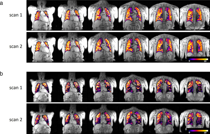
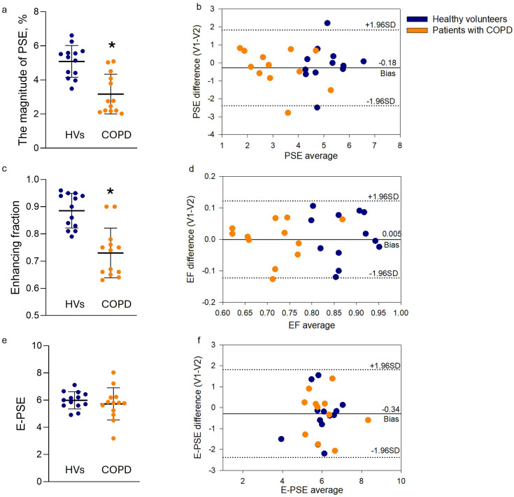
背景:T2*加权氧增强MRI (T2*-OE-MRI)可以直接评估肺通气,将氧气作为吸入示踪气体。它已在健康志愿者(HVs)和囊性纤维化患者中显示出前景,但尚未在慢性阻塞性肺疾病(COPD)患者中得到证实。研究问题:确定T2*-OE-MRI在COPD患者中的可行性和可重复性。评估肺通气、肺功能测试(pft)和功能限制测量之间的相关性。研究设计和方法:13名轻至重度COPD患者和13名hv患者接受了pft、肺清除率指数(LCI)测量、增量运动试验(仅限患者)和2次3 t肺部MRI扫描。对于T2*- e -MRI,参与者在图像采集期间佩戴非再呼吸面罩并给予100%氧气。结果:患者(年龄:63(55-72)岁,1秒内用力呼气量(FEV1): 63(36-79) %预测,中位数(IQR))有肺气体潴留、小气道疾病(SAD)和通气异质性的证据。在T2*-OE-MRI期间,患者与HVs相比,常氧和高氧时平均信号强度(信号增强百分比(PSE))和增强分数(EF)的百分比差值分别为2.77(2.19-4.19)比5.34(4.33-5.61)%和0.74(0.66-0.77)比0.89 (0.82-0.94),p1、LCI和SAD指数与运动能力、动态过度膨胀和运动时呼吸困难强度的测量相关。解释:在COPD患者中,T2*-OE-MRI是可行且可重复的,并提供与疾病严重程度、功能限制和用力呼吸困难的生理测量相关的肺通气区域信息。
T2*-weighted oxygen-enhanced pulmonary MRI in COPD is linked to resting and exertional functional measurements.
Background: T2*-weighted oxygen-enhanced MRI (T2*-OE-MRI) may directly assess pulmonary ventilation using oxygen as an inhaled tracer gas. It has shown promise in healthy volunteers (HVs) and cystic fibrosis but has yet to be demonstrated in patients with chronic obstructive pulmonary disease (COPD).
Research question: To determine the feasibility and repeatability of T2*-OE-MRI in patients with COPD. To assess correlations between T2*-OE-MRI measurements of pulmonary ventilation, pulmonary function tests (PFTs) and measures of functional limitation.
Study design and methods: 13 patients with mild-to-severe COPD and 13 HVs underwent PFTs, lung clearance index (LCI) measurement, incremental exercise test (patients only) and two lung MRI scans at 3 T. For T2*-OE-MRI, participants were fitted with a non-rebreathing face mask and given 100% oxygen during image acquisition.
Results: Patients (age: 63 (55-72) years, forced expiratory volume in 1 s (FEV1): 63 (36-79) %predicted, median (IQR)) had evidence of pulmonary gas trapping, small airway disease (SAD) and ventilation heterogeneity. During T2*-OE-MRI, the magnitude of the percentage difference between mean signal intensity at normoxia and hyperoxia (percent signal enhancement (PSE)) and the enhancing fraction (EF) were lower in patients versus HVs (2.77 (2.19-4.19) vs 5.34 (4.33-5.61) % and 0.74 (0.66-0.77) vs 0.89 (0.82-0.94), respectively, both p<0.001). Intraclass correlation coefficient values indicated moderate (0.74) and good (0.80) repeatability for PSE and EF, respectively. PSE and EF significantly correlated with FEV1, LCI and SAD indices, and in COPD, they correlated with measures of exercise capacity, dynamic hyperinflation and dyspnoea intensity during exercise.
Interpretation: In patients with COPD, T2*-OE-MRI is feasible and repeatable and provides regional information on pulmonary ventilation that is linked with physiological measures of disease severity, functional limitation and exertional dyspnoea.
期刊介绍:
BMJ Open Respiratory Research is a peer-reviewed, open access journal publishing respiratory and critical care medicine. It is the sister journal to Thorax and co-owned by the British Thoracic Society and BMJ. The journal focuses on robustness of methodology and scientific rigour with less emphasis on novelty or perceived impact. BMJ Open Respiratory Research operates a rapid review process, with continuous publication online, ensuring timely, up-to-date research is available worldwide. The journal publishes review articles and all research study types: Basic science including laboratory based experiments and animal models, Pilot studies or proof of concept, Observational studies, Study protocols, Registries, Clinical trials from phase I to multicentre randomised clinical trials, Systematic reviews and meta-analyses.


 求助内容:
求助内容: 应助结果提醒方式:


