{"title":"成人发病的孤立性颈肌张力障碍是否需要常规神经影像学检查?","authors":"Elina Myller, Oskari Korhonen, Juho Joutsa","doi":"10.5334/tohm.1049","DOIUrl":null,"url":null,"abstract":"<p><strong>Background: </strong>Clinical practices regarding neuroimaging in isolated cervical dystonia vary across countries and there are no published studies investigating the need of routine neuroimaging in this patient population.</p><p><strong>Objectives: </strong>To investigate if structural neuroimaging is needed in patients with isolated cervical dystonia.</p><p><strong>Methods: </strong>Patients with adult-onset cervical dystonia were identified from a systematic search of the medical records of Turku University Hospital 1996-2022. Clinical and structural neuroimaging data were reviewed by the investigators to evaluate the etiology of dystonia, specifically to identify cases of secondary dystonia caused by structural brain abnormalities.</p><p><strong>Results: </strong>365 patients with cervical dystonia without other movement disorders with presumed idiopathic or uncertain etiology prior to brain imaging were identified. 282 (77.3%) were scanned using head MRI or CT. Acquired brain lesions were identified in nine (2.5% of all patients) and were significantly more common in patients with vs. without (i.e. isolated) other neurological features (<i>P</i> < 0.001). Lesions in patients with other neurological features were considered likely (n = 4) or possibly (n = 2) causal, but all lesions in patients with isolated cervical dystonia (n = 3) were considered incidental. None of the patients showed signs of progressive neurodegeneration.</p><p><strong>Conclusions: </strong>Routine neuroimaging is not necessary in patients with adult-onset isolated cervical dystonia.</p><p><strong>Highlights: </strong>Studies investigating the need of structural neuroimaging in isolated, adult-onset cervical dystonia are scarce and opinions on this issue are divided among experts.In this study, we reviewed clinical and imaging data of all patients with cervical dystonia with presumed idiopathic or uncertain etiology prior to brain imaging treated at a regional tertiary care hospital between 1996-2022 to investigate the yield of structural brain imaging in these patients.Of the included 365 patients, none showed evidence of progressive neurodegeneration underlying the symptoms and only six (1.6%) showed acquired brain lesions that were considered possibly or likely causal for cervical dystonia.All the six patients with possible or likely lesion-induced cervical dystonia showed cervical dystonia combined with other neurological features, indicating that routine neuroimaging is not needed in isolated, adult-onset cervical dystonia.</p>","PeriodicalId":23317,"journal":{"name":"Tremor and Other Hyperkinetic Movements","volume":"15 ","pages":"36"},"PeriodicalIF":2.1000,"publicationDate":"2025-08-06","publicationTypes":"Journal Article","fieldsOfStudy":null,"isOpenAccess":false,"openAccessPdf":"https://www.ncbi.nlm.nih.gov/pmc/articles/PMC12330805/pdf/","citationCount":"0","resultStr":"{\"title\":\"Is Routine Neuroimaging Needed in Adult-Onset Isolated Cervical Dystonia?\",\"authors\":\"Elina Myller, Oskari Korhonen, Juho Joutsa\",\"doi\":\"10.5334/tohm.1049\",\"DOIUrl\":null,\"url\":null,\"abstract\":\"<p><strong>Background: </strong>Clinical practices regarding neuroimaging in isolated cervical dystonia vary across countries and there are no published studies investigating the need of routine neuroimaging in this patient population.</p><p><strong>Objectives: </strong>To investigate if structural neuroimaging is needed in patients with isolated cervical dystonia.</p><p><strong>Methods: </strong>Patients with adult-onset cervical dystonia were identified from a systematic search of the medical records of Turku University Hospital 1996-2022. Clinical and structural neuroimaging data were reviewed by the investigators to evaluate the etiology of dystonia, specifically to identify cases of secondary dystonia caused by structural brain abnormalities.</p><p><strong>Results: </strong>365 patients with cervical dystonia without other movement disorders with presumed idiopathic or uncertain etiology prior to brain imaging were identified. 282 (77.3%) were scanned using head MRI or CT. Acquired brain lesions were identified in nine (2.5% of all patients) and were significantly more common in patients with vs. without (i.e. isolated) other neurological features (<i>P</i> < 0.001). Lesions in patients with other neurological features were considered likely (n = 4) or possibly (n = 2) causal, but all lesions in patients with isolated cervical dystonia (n = 3) were considered incidental. None of the patients showed signs of progressive neurodegeneration.</p><p><strong>Conclusions: </strong>Routine neuroimaging is not necessary in patients with adult-onset isolated cervical dystonia.</p><p><strong>Highlights: </strong>Studies investigating the need of structural neuroimaging in isolated, adult-onset cervical dystonia are scarce and opinions on this issue are divided among experts.In this study, we reviewed clinical and imaging data of all patients with cervical dystonia with presumed idiopathic or uncertain etiology prior to brain imaging treated at a regional tertiary care hospital between 1996-2022 to investigate the yield of structural brain imaging in these patients.Of the included 365 patients, none showed evidence of progressive neurodegeneration underlying the symptoms and only six (1.6%) showed acquired brain lesions that were considered possibly or likely causal for cervical dystonia.All the six patients with possible or likely lesion-induced cervical dystonia showed cervical dystonia combined with other neurological features, indicating that routine neuroimaging is not needed in isolated, adult-onset cervical dystonia.</p>\",\"PeriodicalId\":23317,\"journal\":{\"name\":\"Tremor and Other Hyperkinetic Movements\",\"volume\":\"15 \",\"pages\":\"36\"},\"PeriodicalIF\":2.1000,\"publicationDate\":\"2025-08-06\",\"publicationTypes\":\"Journal Article\",\"fieldsOfStudy\":null,\"isOpenAccess\":false,\"openAccessPdf\":\"https://www.ncbi.nlm.nih.gov/pmc/articles/PMC12330805/pdf/\",\"citationCount\":\"0\",\"resultStr\":null,\"platform\":\"Semanticscholar\",\"paperid\":null,\"PeriodicalName\":\"Tremor and Other Hyperkinetic Movements\",\"FirstCategoryId\":\"1085\",\"ListUrlMain\":\"https://doi.org/10.5334/tohm.1049\",\"RegionNum\":0,\"RegionCategory\":null,\"ArticlePicture\":[],\"TitleCN\":null,\"AbstractTextCN\":null,\"PMCID\":null,\"EPubDate\":\"2025/1/1 0:00:00\",\"PubModel\":\"eCollection\",\"JCR\":\"Q2\",\"JCRName\":\"CLINICAL NEUROLOGY\",\"Score\":null,\"Total\":0}","platform":"Semanticscholar","paperid":null,"PeriodicalName":"Tremor and Other Hyperkinetic Movements","FirstCategoryId":"1085","ListUrlMain":"https://doi.org/10.5334/tohm.1049","RegionNum":0,"RegionCategory":null,"ArticlePicture":[],"TitleCN":null,"AbstractTextCN":null,"PMCID":null,"EPubDate":"2025/1/1 0:00:00","PubModel":"eCollection","JCR":"Q2","JCRName":"CLINICAL NEUROLOGY","Score":null,"Total":0}
Is Routine Neuroimaging Needed in Adult-Onset Isolated Cervical Dystonia?
Background: Clinical practices regarding neuroimaging in isolated cervical dystonia vary across countries and there are no published studies investigating the need of routine neuroimaging in this patient population.
Objectives: To investigate if structural neuroimaging is needed in patients with isolated cervical dystonia.
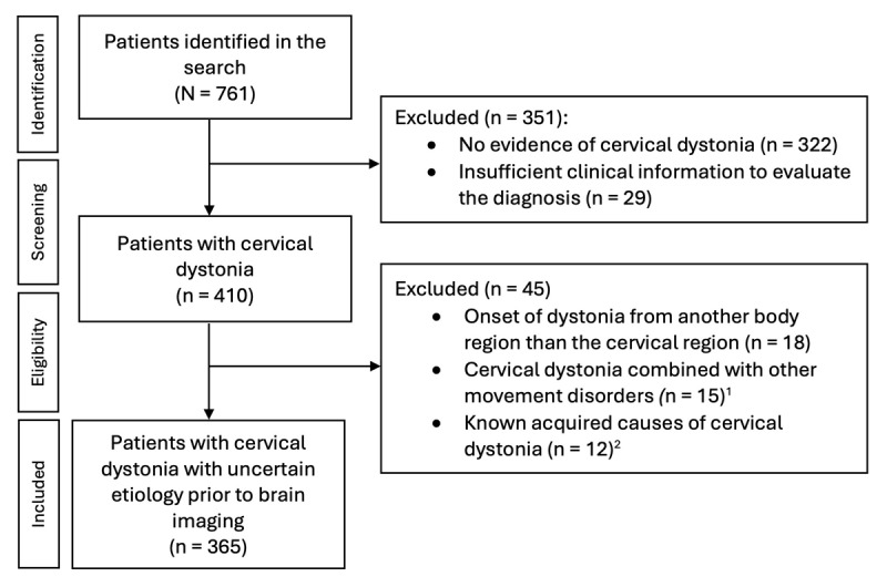
Methods: Patients with adult-onset cervical dystonia were identified from a systematic search of the medical records of Turku University Hospital 1996-2022. Clinical and structural neuroimaging data were reviewed by the investigators to evaluate the etiology of dystonia, specifically to identify cases of secondary dystonia caused by structural brain abnormalities.
Results: 365 patients with cervical dystonia without other movement disorders with presumed idiopathic or uncertain etiology prior to brain imaging were identified. 282 (77.3%) were scanned using head MRI or CT. Acquired brain lesions were identified in nine (2.5% of all patients) and were significantly more common in patients with vs. without (i.e. isolated) other neurological features (P < 0.001). Lesions in patients with other neurological features were considered likely (n = 4) or possibly (n = 2) causal, but all lesions in patients with isolated cervical dystonia (n = 3) were considered incidental. None of the patients showed signs of progressive neurodegeneration.
Conclusions: Routine neuroimaging is not necessary in patients with adult-onset isolated cervical dystonia.
Highlights: Studies investigating the need of structural neuroimaging in isolated, adult-onset cervical dystonia are scarce and opinions on this issue are divided among experts.In this study, we reviewed clinical and imaging data of all patients with cervical dystonia with presumed idiopathic or uncertain etiology prior to brain imaging treated at a regional tertiary care hospital between 1996-2022 to investigate the yield of structural brain imaging in these patients.Of the included 365 patients, none showed evidence of progressive neurodegeneration underlying the symptoms and only six (1.6%) showed acquired brain lesions that were considered possibly or likely causal for cervical dystonia.All the six patients with possible or likely lesion-induced cervical dystonia showed cervical dystonia combined with other neurological features, indicating that routine neuroimaging is not needed in isolated, adult-onset cervical dystonia.


 求助内容:
求助内容: 应助结果提醒方式:


