Akos Horvath, Rita Csercsik, Janos Norbert Gyebnar, Daniel Sandor Veres, Pal Maurovich-Horvat, Akos Menyhart, Viktoria Gyergyoi, Peter Vince Balint, Nikolett Marton
{"title":"优化光子计数CT造影剂时机评估周围性关节炎。","authors":"Akos Horvath, Rita Csercsik, Janos Norbert Gyebnar, Daniel Sandor Veres, Pal Maurovich-Horvat, Akos Menyhart, Viktoria Gyergyoi, Peter Vince Balint, Nikolett Marton","doi":"10.1007/s00256-025-04993-4","DOIUrl":null,"url":null,"abstract":"<p><strong>Objective: </strong>Photon-counting detector CT (PCD-CT) allows for iodine mapping of inflamed tissues in peripheral immune-mediated arthritis, supporting diagnosis and disease activity assessment. This study aims to identify the optimal timing for image acquisition after intravenous iodinated contrast administration to maximize enhancement and contrast with surrounding tissues.</p><p><strong>Methods: </strong>High-resolution PCD-CT scans of bilateral wrist-hand regions were obtained from 26 patients with peripheral arthritis, both native and post-contrast (1 ml/kg intravenous iodinated contrast at 2.5 ml/sec flow) at 120-, 180-, and 240-s delay phases. Iodine maps were constructed from spectral data. Phases were compared based on densities and iodine concentrations measured in synovial, tenosynovial, and periungual tissues, with muscle, fat, and vessels as controls. We used descriptive statistics and mixed-effects linear regression inferential models for the comparisons. Synovitis and tenosynovitis were verified by ultrasound measurements.</p><p><strong>Results: </strong>No significant differences (p > 0.05) were found in iodine concentration or density across the 120-, 180-, and 240-s post-contrast phases in inflamed synovial, tenosynovial, and periungual soft tissues. Inflamed tissues showed significant and consistent differences in iodine concentration from muscle and fat (p < 0.0001) across all phases, while the greatest differentiation from vessels was in the 120-s phase. The effective dose was identical across all post-contrast phases (0.028 ± 0.0035 mSv).</p><p><strong>Conclusion: </strong>Iodine uptake in inflamed tissues was identical across all three post-contrast phases. However, the 120-s phase offered the highest contrast between inflammation and surrounding vascular structures while minimizing scan time, supporting its use for standardized follow-up imaging.</p>","PeriodicalId":21783,"journal":{"name":"Skeletal Radiology","volume":" ","pages":"2565-2578"},"PeriodicalIF":2.2000,"publicationDate":"2025-11-01","publicationTypes":"Journal Article","fieldsOfStudy":null,"isOpenAccess":false,"openAccessPdf":"https://www.ncbi.nlm.nih.gov/pmc/articles/PMC12460460/pdf/","citationCount":"0","resultStr":"{\"title\":\"Optimizing contrast timing in photon-counting detector CT for the assessment of peripheral arthritis.\",\"authors\":\"Akos Horvath, Rita Csercsik, Janos Norbert Gyebnar, Daniel Sandor Veres, Pal Maurovich-Horvat, Akos Menyhart, Viktoria Gyergyoi, Peter Vince Balint, Nikolett Marton\",\"doi\":\"10.1007/s00256-025-04993-4\",\"DOIUrl\":null,\"url\":null,\"abstract\":\"<p><strong>Objective: </strong>Photon-counting detector CT (PCD-CT) allows for iodine mapping of inflamed tissues in peripheral immune-mediated arthritis, supporting diagnosis and disease activity assessment. This study aims to identify the optimal timing for image acquisition after intravenous iodinated contrast administration to maximize enhancement and contrast with surrounding tissues.</p><p><strong>Methods: </strong>High-resolution PCD-CT scans of bilateral wrist-hand regions were obtained from 26 patients with peripheral arthritis, both native and post-contrast (1 ml/kg intravenous iodinated contrast at 2.5 ml/sec flow) at 120-, 180-, and 240-s delay phases. Iodine maps were constructed from spectral data. Phases were compared based on densities and iodine concentrations measured in synovial, tenosynovial, and periungual tissues, with muscle, fat, and vessels as controls. We used descriptive statistics and mixed-effects linear regression inferential models for the comparisons. Synovitis and tenosynovitis were verified by ultrasound measurements.</p><p><strong>Results: </strong>No significant differences (p > 0.05) were found in iodine concentration or density across the 120-, 180-, and 240-s post-contrast phases in inflamed synovial, tenosynovial, and periungual soft tissues. Inflamed tissues showed significant and consistent differences in iodine concentration from muscle and fat (p < 0.0001) across all phases, while the greatest differentiation from vessels was in the 120-s phase. The effective dose was identical across all post-contrast phases (0.028 ± 0.0035 mSv).</p><p><strong>Conclusion: </strong>Iodine uptake in inflamed tissues was identical across all three post-contrast phases. However, the 120-s phase offered the highest contrast between inflammation and surrounding vascular structures while minimizing scan time, supporting its use for standardized follow-up imaging.</p>\",\"PeriodicalId\":21783,\"journal\":{\"name\":\"Skeletal Radiology\",\"volume\":\" \",\"pages\":\"2565-2578\"},\"PeriodicalIF\":2.2000,\"publicationDate\":\"2025-11-01\",\"publicationTypes\":\"Journal Article\",\"fieldsOfStudy\":null,\"isOpenAccess\":false,\"openAccessPdf\":\"https://www.ncbi.nlm.nih.gov/pmc/articles/PMC12460460/pdf/\",\"citationCount\":\"0\",\"resultStr\":null,\"platform\":\"Semanticscholar\",\"paperid\":null,\"PeriodicalName\":\"Skeletal Radiology\",\"FirstCategoryId\":\"3\",\"ListUrlMain\":\"https://doi.org/10.1007/s00256-025-04993-4\",\"RegionNum\":3,\"RegionCategory\":\"医学\",\"ArticlePicture\":[],\"TitleCN\":null,\"AbstractTextCN\":null,\"PMCID\":null,\"EPubDate\":\"2025/8/8 0:00:00\",\"PubModel\":\"Epub\",\"JCR\":\"Q2\",\"JCRName\":\"ORTHOPEDICS\",\"Score\":null,\"Total\":0}","platform":"Semanticscholar","paperid":null,"PeriodicalName":"Skeletal Radiology","FirstCategoryId":"3","ListUrlMain":"https://doi.org/10.1007/s00256-025-04993-4","RegionNum":3,"RegionCategory":"医学","ArticlePicture":[],"TitleCN":null,"AbstractTextCN":null,"PMCID":null,"EPubDate":"2025/8/8 0:00:00","PubModel":"Epub","JCR":"Q2","JCRName":"ORTHOPEDICS","Score":null,"Total":0}
Optimizing contrast timing in photon-counting detector CT for the assessment of peripheral arthritis.
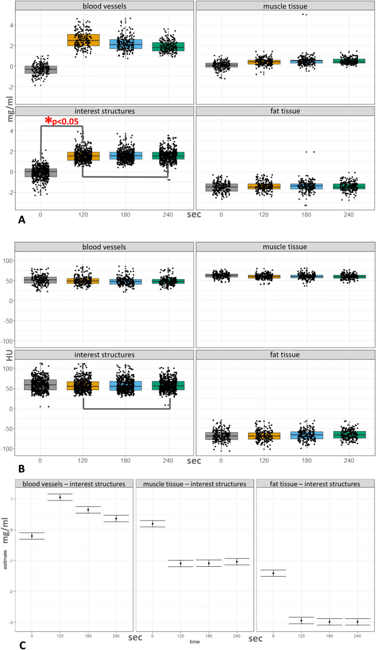
Objective: Photon-counting detector CT (PCD-CT) allows for iodine mapping of inflamed tissues in peripheral immune-mediated arthritis, supporting diagnosis and disease activity assessment. This study aims to identify the optimal timing for image acquisition after intravenous iodinated contrast administration to maximize enhancement and contrast with surrounding tissues.
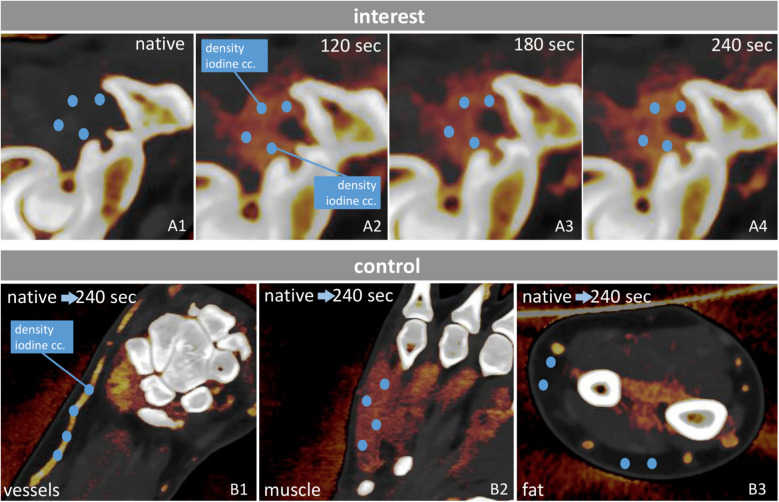
Methods: High-resolution PCD-CT scans of bilateral wrist-hand regions were obtained from 26 patients with peripheral arthritis, both native and post-contrast (1 ml/kg intravenous iodinated contrast at 2.5 ml/sec flow) at 120-, 180-, and 240-s delay phases. Iodine maps were constructed from spectral data. Phases were compared based on densities and iodine concentrations measured in synovial, tenosynovial, and periungual tissues, with muscle, fat, and vessels as controls. We used descriptive statistics and mixed-effects linear regression inferential models for the comparisons. Synovitis and tenosynovitis were verified by ultrasound measurements.
Results: No significant differences (p > 0.05) were found in iodine concentration or density across the 120-, 180-, and 240-s post-contrast phases in inflamed synovial, tenosynovial, and periungual soft tissues. Inflamed tissues showed significant and consistent differences in iodine concentration from muscle and fat (p < 0.0001) across all phases, while the greatest differentiation from vessels was in the 120-s phase. The effective dose was identical across all post-contrast phases (0.028 ± 0.0035 mSv).
Conclusion: Iodine uptake in inflamed tissues was identical across all three post-contrast phases. However, the 120-s phase offered the highest contrast between inflammation and surrounding vascular structures while minimizing scan time, supporting its use for standardized follow-up imaging.
期刊介绍:
Skeletal Radiology provides a forum for the dissemination of current knowledge and information dealing with disorders of the musculoskeletal system including the spine. While emphasizing the radiological aspects of the many varied skeletal abnormalities, the journal also adopts an interdisciplinary approach, reflecting the membership of the International Skeletal Society. Thus, the anatomical, pathological, physiological, clinical, metabolic and epidemiological aspects of the many entities affecting the skeleton receive appropriate consideration.
This is the Journal of the International Skeletal Society and the Official Journal of the Society of Skeletal Radiology and the Australasian Musculoskelelal Imaging Group.


 求助内容:
求助内容: 应助结果提醒方式:


