{"title":"颌骨破坏性病变、牙龈内阿米巴和放线菌合并感染的刮刮细胞学应用前景:回顾性分析。","authors":"Tsuyoshi Kurobane, Sukhbaatar Ariunbuyan, Shiro Mori, Hiroyuki Kumamoto, Tsuyoshi Sugiura","doi":"10.1186/s13005-025-00535-4","DOIUrl":null,"url":null,"abstract":"<p><strong>Objectives: </strong>Microbial infections can influence destructive jawbone lesions, yet the role of Entamoeba gingivalis (E. gingivalis) in these lesions is poorly understood, particularly when co-infected with Actinomyces. This study introduces a novel clinicopathological perspective on jawbone destruction linked to E. gingivalis and Actinomyces co-infection, aiming to improve diagnosis and treatment. In this study, we tried to evaluate minimally invasive methods for improving diagnosis and treatment for jawbone destruction, possibly linked to E. gingivalis and Actinomyces co-infection. We analyzed 11 cases of jawbone destructive lesions observed in patients (ages 42 to 83, both female and male) who visited Tohoku University Hospital from January 2015 to December 2021, in which scraping cytology detected E. gingivalis and Actinomyces.</p><p><strong>Materials and methods: </strong>We thoroughly assessed clinical symptoms, imaging findings, and treatment outcomes, including the duration of antimicrobial therapy, to identify distinctive patterns associated with this co-infection.</p><p><strong>Results: </strong>On imaging, patients exhibited significant mandibular resorption and sclerosis, despite the absence of typical symptoms of osteomyelitis or actinomycosis. Notably, E. gingivalis alone was linked to faster wound healing and shorter antimicrobial therapy durations compared to cases with concurrent Actinomyces infection (p = 0.011). The use of minimally invasive procedures, focusing on removing necrotic bone and loose teeth, led to effective healing and rapid recovery across all cases, offering promising prospects for enhanced treatment outcomes.</p><p><strong>Conclusion: </strong>This study underscores the value of scraping cytology for early identification of E. gingivalis and Actinomyces in jawbone lesions. Early detection and targeted intervention can prevent disease progression, enhance patient outcomes, and optimize antimicrobial use.</p><p><strong>Clinical relevance: </strong>These findings highlight the need for increased awareness of microbial co-infections in jaw osteomyelitis and support a new clinicopathological approach to diagnosis and management.</p>","PeriodicalId":12994,"journal":{"name":"Head & Face Medicine","volume":"21 1","pages":"58"},"PeriodicalIF":2.4000,"publicationDate":"2025-08-07","publicationTypes":"Journal Article","fieldsOfStudy":null,"isOpenAccess":false,"openAccessPdf":"https://www.ncbi.nlm.nih.gov/pmc/articles/PMC12329943/pdf/","citationCount":"0","resultStr":"{\"title\":\"Perspective on the use of scraping cytology for jawbone destructive lesions, entamoeba gingivalis, and actinomyces co-infection: a retrospective analysis.\",\"authors\":\"Tsuyoshi Kurobane, Sukhbaatar Ariunbuyan, Shiro Mori, Hiroyuki Kumamoto, Tsuyoshi Sugiura\",\"doi\":\"10.1186/s13005-025-00535-4\",\"DOIUrl\":null,\"url\":null,\"abstract\":\"<p><strong>Objectives: </strong>Microbial infections can influence destructive jawbone lesions, yet the role of Entamoeba gingivalis (E. gingivalis) in these lesions is poorly understood, particularly when co-infected with Actinomyces. This study introduces a novel clinicopathological perspective on jawbone destruction linked to E. gingivalis and Actinomyces co-infection, aiming to improve diagnosis and treatment. In this study, we tried to evaluate minimally invasive methods for improving diagnosis and treatment for jawbone destruction, possibly linked to E. gingivalis and Actinomyces co-infection. We analyzed 11 cases of jawbone destructive lesions observed in patients (ages 42 to 83, both female and male) who visited Tohoku University Hospital from January 2015 to December 2021, in which scraping cytology detected E. gingivalis and Actinomyces.</p><p><strong>Materials and methods: </strong>We thoroughly assessed clinical symptoms, imaging findings, and treatment outcomes, including the duration of antimicrobial therapy, to identify distinctive patterns associated with this co-infection.</p><p><strong>Results: </strong>On imaging, patients exhibited significant mandibular resorption and sclerosis, despite the absence of typical symptoms of osteomyelitis or actinomycosis. Notably, E. gingivalis alone was linked to faster wound healing and shorter antimicrobial therapy durations compared to cases with concurrent Actinomyces infection (p = 0.011). The use of minimally invasive procedures, focusing on removing necrotic bone and loose teeth, led to effective healing and rapid recovery across all cases, offering promising prospects for enhanced treatment outcomes.</p><p><strong>Conclusion: </strong>This study underscores the value of scraping cytology for early identification of E. gingivalis and Actinomyces in jawbone lesions. Early detection and targeted intervention can prevent disease progression, enhance patient outcomes, and optimize antimicrobial use.</p><p><strong>Clinical relevance: </strong>These findings highlight the need for increased awareness of microbial co-infections in jaw osteomyelitis and support a new clinicopathological approach to diagnosis and management.</p>\",\"PeriodicalId\":12994,\"journal\":{\"name\":\"Head & Face Medicine\",\"volume\":\"21 1\",\"pages\":\"58\"},\"PeriodicalIF\":2.4000,\"publicationDate\":\"2025-08-07\",\"publicationTypes\":\"Journal Article\",\"fieldsOfStudy\":null,\"isOpenAccess\":false,\"openAccessPdf\":\"https://www.ncbi.nlm.nih.gov/pmc/articles/PMC12329943/pdf/\",\"citationCount\":\"0\",\"resultStr\":null,\"platform\":\"Semanticscholar\",\"paperid\":null,\"PeriodicalName\":\"Head & Face Medicine\",\"FirstCategoryId\":\"3\",\"ListUrlMain\":\"https://doi.org/10.1186/s13005-025-00535-4\",\"RegionNum\":2,\"RegionCategory\":\"医学\",\"ArticlePicture\":[],\"TitleCN\":null,\"AbstractTextCN\":null,\"PMCID\":null,\"EPubDate\":\"\",\"PubModel\":\"\",\"JCR\":\"Q2\",\"JCRName\":\"DENTISTRY, ORAL SURGERY & MEDICINE\",\"Score\":null,\"Total\":0}","platform":"Semanticscholar","paperid":null,"PeriodicalName":"Head & Face Medicine","FirstCategoryId":"3","ListUrlMain":"https://doi.org/10.1186/s13005-025-00535-4","RegionNum":2,"RegionCategory":"医学","ArticlePicture":[],"TitleCN":null,"AbstractTextCN":null,"PMCID":null,"EPubDate":"","PubModel":"","JCR":"Q2","JCRName":"DENTISTRY, ORAL SURGERY & MEDICINE","Score":null,"Total":0}
Perspective on the use of scraping cytology for jawbone destructive lesions, entamoeba gingivalis, and actinomyces co-infection: a retrospective analysis.
Objectives: Microbial infections can influence destructive jawbone lesions, yet the role of Entamoeba gingivalis (E. gingivalis) in these lesions is poorly understood, particularly when co-infected with Actinomyces. This study introduces a novel clinicopathological perspective on jawbone destruction linked to E. gingivalis and Actinomyces co-infection, aiming to improve diagnosis and treatment. In this study, we tried to evaluate minimally invasive methods for improving diagnosis and treatment for jawbone destruction, possibly linked to E. gingivalis and Actinomyces co-infection. We analyzed 11 cases of jawbone destructive lesions observed in patients (ages 42 to 83, both female and male) who visited Tohoku University Hospital from January 2015 to December 2021, in which scraping cytology detected E. gingivalis and Actinomyces.
Materials and methods: We thoroughly assessed clinical symptoms, imaging findings, and treatment outcomes, including the duration of antimicrobial therapy, to identify distinctive patterns associated with this co-infection.
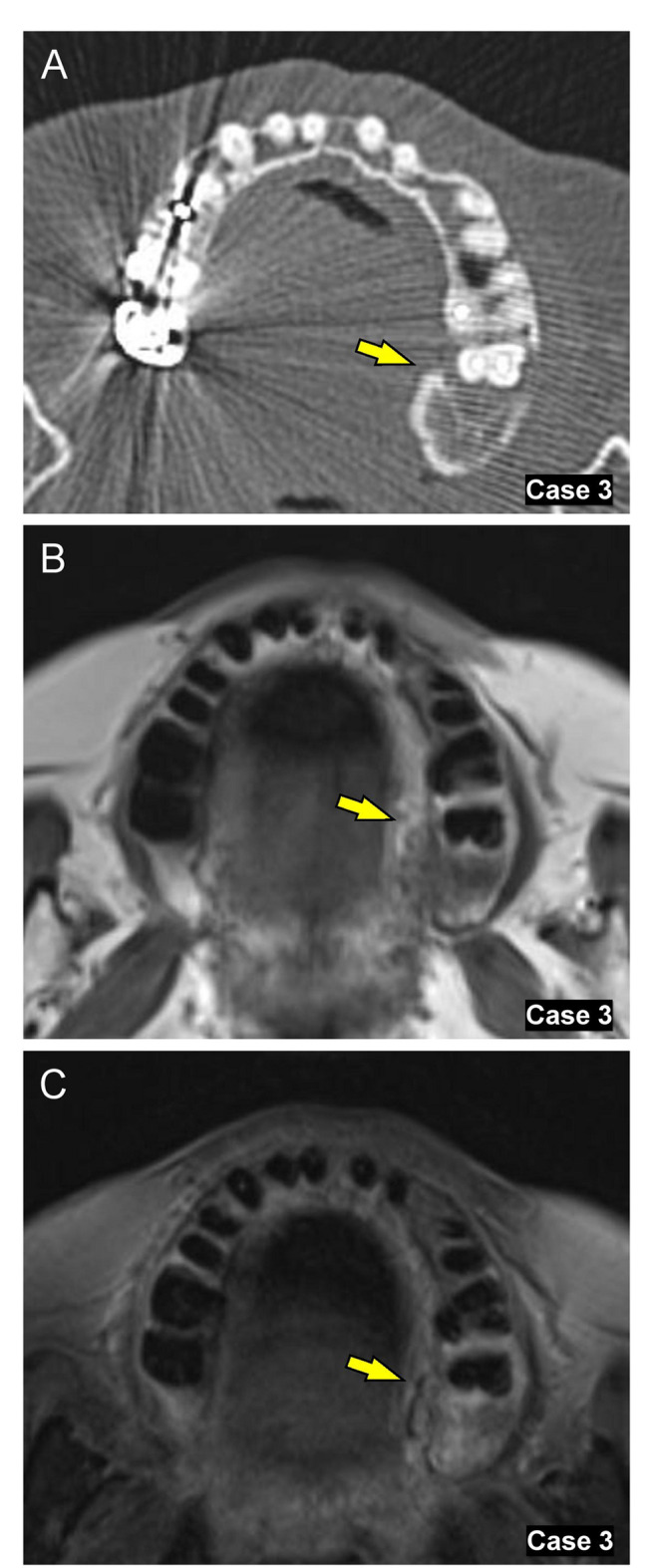
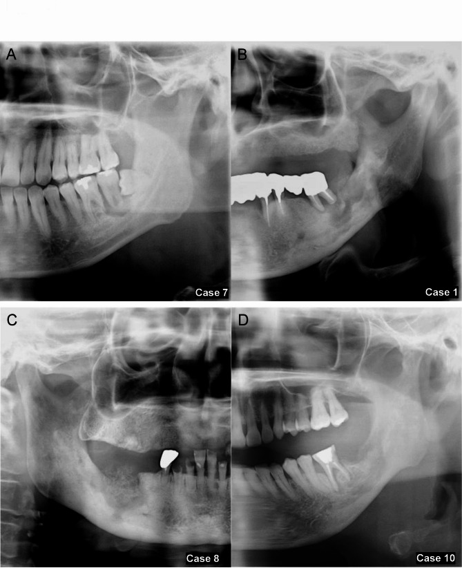
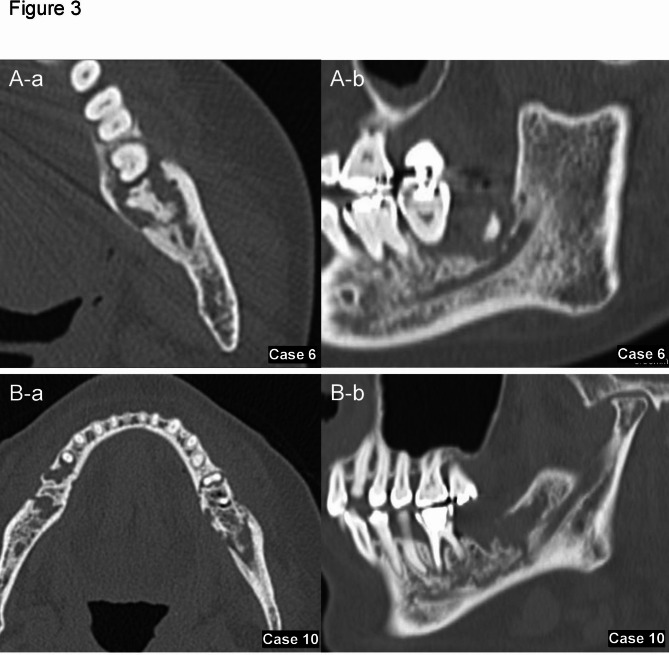
Results: On imaging, patients exhibited significant mandibular resorption and sclerosis, despite the absence of typical symptoms of osteomyelitis or actinomycosis. Notably, E. gingivalis alone was linked to faster wound healing and shorter antimicrobial therapy durations compared to cases with concurrent Actinomyces infection (p = 0.011). The use of minimally invasive procedures, focusing on removing necrotic bone and loose teeth, led to effective healing and rapid recovery across all cases, offering promising prospects for enhanced treatment outcomes.
Conclusion: This study underscores the value of scraping cytology for early identification of E. gingivalis and Actinomyces in jawbone lesions. Early detection and targeted intervention can prevent disease progression, enhance patient outcomes, and optimize antimicrobial use.
Clinical relevance: These findings highlight the need for increased awareness of microbial co-infections in jaw osteomyelitis and support a new clinicopathological approach to diagnosis and management.
期刊介绍:
Head & Face Medicine is a multidisciplinary open access journal that publishes basic and clinical research concerning all aspects of cranial, facial and oral conditions.
The journal covers all aspects of cranial, facial and oral diseases and their management. It has been designed as a multidisciplinary journal for clinicians and researchers involved in the diagnostic and therapeutic aspects of diseases which affect the human head and face. The journal is wide-ranging, covering the development, aetiology, epidemiology and therapy of head and face diseases to the basic science that underlies these diseases. Management of head and face diseases includes all aspects of surgical and non-surgical treatments including psychopharmacological therapies.
 求助内容:
求助内容: 应助结果提醒方式:


