Thai Hau Koo, Venkata Sunkesula, Salah Abdel Jalil, Richard Wong, Ala Abdel-Jalil, Elham Abdel Jalil
{"title":"腺泡细胞囊腺瘤合并胰腺上皮内瘤变1例:是否总是良性的?","authors":"Thai Hau Koo, Venkata Sunkesula, Salah Abdel Jalil, Richard Wong, Ala Abdel-Jalil, Elham Abdel Jalil","doi":"10.1159/000546668","DOIUrl":null,"url":null,"abstract":"<p><strong>Introduction: </strong>The exact etiology of acinar cell cystadenoma (ACC) has been debated, primarily whether it originates from or carries the risk of an underlying neoplasia. Pancreatic intraepithelial neoplasia (PanIN) is presumed to be a noninvasive precursor of pancreatic ductal adenocarcinoma. This report presents a rare case of ACC with low-grade PanIN that required surgical resection.</p><p><strong>Case presentation: </strong>A 60-year-old female with an unremarkable medical history presented with epigastric pain for 2 weeks. Her initial laboratory workup was notable for mild isolated elevation of alkaline phosphatase. Abdominal computed tomography revealed a 5.0 × 4.0 cm cystic lesion in the pancreatic head with thick internal septations. Magnetic resonance cholangiopancreatography showed a 5.2 × 4.5 × 6.8 cm lobulated cystic lesion in the pancreatic head with a microcystic configuration, multiple internal septations, and a hypointense central scar. Endosonographic examination showed a large multicystic lesion in the pancreatic head region. Fine-needle aspiration showed a carcinoembryonic antigen level of 555 ng/mL and an amylase level of 13,593 U/L. No KRAS or GNAS mutations or loss of heterozygosity was detected. Subsequently, the patient underwent a Whipple procedure. Pathologic examination revealed a complex cystic lesion with well-differentiated acinar cells and patches of ductal epithelium compatible with ACC. Histological examination confirmed the presence of low-grade PanIN without invasive carcinoma. The patient recovered well from surgery, and repeat imaging 2 months later was unremarkable.</p><p><strong>Conclusion: </strong>ACC is a rare benign pancreatic lesion. Low-grade PanIN is typically found in benign pancreatic lesions. Resection is recommended for symptomatic patients.</p>","PeriodicalId":9614,"journal":{"name":"Case Reports in Gastroenterology","volume":"19 1","pages":"534-540"},"PeriodicalIF":0.6000,"publicationDate":"2025-08-06","publicationTypes":"Journal Article","fieldsOfStudy":null,"isOpenAccess":false,"openAccessPdf":"https://www.ncbi.nlm.nih.gov/pmc/articles/PMC12327933/pdf/","citationCount":"0","resultStr":"{\"title\":\"A Case Report of Acinar Cell Cystadenoma with Pancreatic Intraepithelial Neoplasia: Is It Always Benign?\",\"authors\":\"Thai Hau Koo, Venkata Sunkesula, Salah Abdel Jalil, Richard Wong, Ala Abdel-Jalil, Elham Abdel Jalil\",\"doi\":\"10.1159/000546668\",\"DOIUrl\":null,\"url\":null,\"abstract\":\"<p><strong>Introduction: </strong>The exact etiology of acinar cell cystadenoma (ACC) has been debated, primarily whether it originates from or carries the risk of an underlying neoplasia. Pancreatic intraepithelial neoplasia (PanIN) is presumed to be a noninvasive precursor of pancreatic ductal adenocarcinoma. This report presents a rare case of ACC with low-grade PanIN that required surgical resection.</p><p><strong>Case presentation: </strong>A 60-year-old female with an unremarkable medical history presented with epigastric pain for 2 weeks. Her initial laboratory workup was notable for mild isolated elevation of alkaline phosphatase. Abdominal computed tomography revealed a 5.0 × 4.0 cm cystic lesion in the pancreatic head with thick internal septations. Magnetic resonance cholangiopancreatography showed a 5.2 × 4.5 × 6.8 cm lobulated cystic lesion in the pancreatic head with a microcystic configuration, multiple internal septations, and a hypointense central scar. Endosonographic examination showed a large multicystic lesion in the pancreatic head region. Fine-needle aspiration showed a carcinoembryonic antigen level of 555 ng/mL and an amylase level of 13,593 U/L. No KRAS or GNAS mutations or loss of heterozygosity was detected. Subsequently, the patient underwent a Whipple procedure. Pathologic examination revealed a complex cystic lesion with well-differentiated acinar cells and patches of ductal epithelium compatible with ACC. Histological examination confirmed the presence of low-grade PanIN without invasive carcinoma. The patient recovered well from surgery, and repeat imaging 2 months later was unremarkable.</p><p><strong>Conclusion: </strong>ACC is a rare benign pancreatic lesion. Low-grade PanIN is typically found in benign pancreatic lesions. Resection is recommended for symptomatic patients.</p>\",\"PeriodicalId\":9614,\"journal\":{\"name\":\"Case Reports in Gastroenterology\",\"volume\":\"19 1\",\"pages\":\"534-540\"},\"PeriodicalIF\":0.6000,\"publicationDate\":\"2025-08-06\",\"publicationTypes\":\"Journal Article\",\"fieldsOfStudy\":null,\"isOpenAccess\":false,\"openAccessPdf\":\"https://www.ncbi.nlm.nih.gov/pmc/articles/PMC12327933/pdf/\",\"citationCount\":\"0\",\"resultStr\":null,\"platform\":\"Semanticscholar\",\"paperid\":null,\"PeriodicalName\":\"Case Reports in Gastroenterology\",\"FirstCategoryId\":\"1085\",\"ListUrlMain\":\"https://doi.org/10.1159/000546668\",\"RegionNum\":0,\"RegionCategory\":null,\"ArticlePicture\":[],\"TitleCN\":null,\"AbstractTextCN\":null,\"PMCID\":null,\"EPubDate\":\"2025/1/1 0:00:00\",\"PubModel\":\"eCollection\",\"JCR\":\"Q4\",\"JCRName\":\"GASTROENTEROLOGY & HEPATOLOGY\",\"Score\":null,\"Total\":0}","platform":"Semanticscholar","paperid":null,"PeriodicalName":"Case Reports in Gastroenterology","FirstCategoryId":"1085","ListUrlMain":"https://doi.org/10.1159/000546668","RegionNum":0,"RegionCategory":null,"ArticlePicture":[],"TitleCN":null,"AbstractTextCN":null,"PMCID":null,"EPubDate":"2025/1/1 0:00:00","PubModel":"eCollection","JCR":"Q4","JCRName":"GASTROENTEROLOGY & HEPATOLOGY","Score":null,"Total":0}
A Case Report of Acinar Cell Cystadenoma with Pancreatic Intraepithelial Neoplasia: Is It Always Benign?
Introduction: The exact etiology of acinar cell cystadenoma (ACC) has been debated, primarily whether it originates from or carries the risk of an underlying neoplasia. Pancreatic intraepithelial neoplasia (PanIN) is presumed to be a noninvasive precursor of pancreatic ductal adenocarcinoma. This report presents a rare case of ACC with low-grade PanIN that required surgical resection.
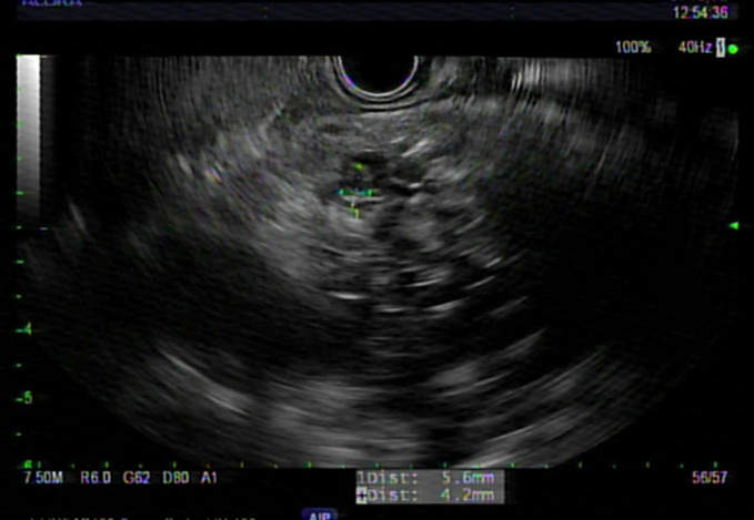
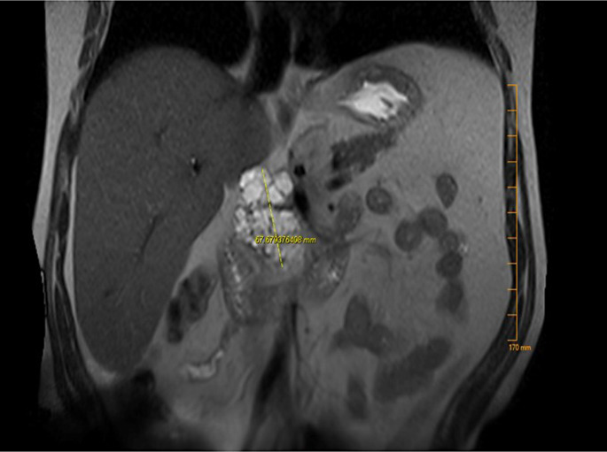
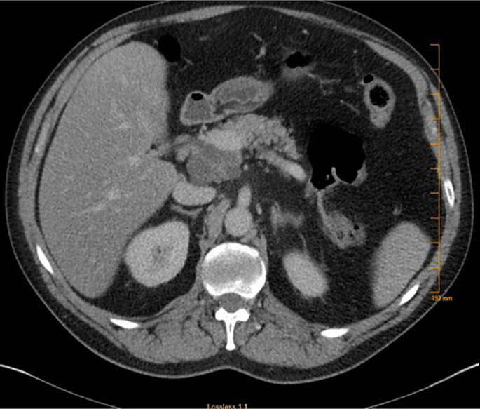
Case presentation: A 60-year-old female with an unremarkable medical history presented with epigastric pain for 2 weeks. Her initial laboratory workup was notable for mild isolated elevation of alkaline phosphatase. Abdominal computed tomography revealed a 5.0 × 4.0 cm cystic lesion in the pancreatic head with thick internal septations. Magnetic resonance cholangiopancreatography showed a 5.2 × 4.5 × 6.8 cm lobulated cystic lesion in the pancreatic head with a microcystic configuration, multiple internal septations, and a hypointense central scar. Endosonographic examination showed a large multicystic lesion in the pancreatic head region. Fine-needle aspiration showed a carcinoembryonic antigen level of 555 ng/mL and an amylase level of 13,593 U/L. No KRAS or GNAS mutations or loss of heterozygosity was detected. Subsequently, the patient underwent a Whipple procedure. Pathologic examination revealed a complex cystic lesion with well-differentiated acinar cells and patches of ductal epithelium compatible with ACC. Histological examination confirmed the presence of low-grade PanIN without invasive carcinoma. The patient recovered well from surgery, and repeat imaging 2 months later was unremarkable.
Conclusion: ACC is a rare benign pancreatic lesion. Low-grade PanIN is typically found in benign pancreatic lesions. Resection is recommended for symptomatic patients.

 求助内容:
求助内容: 应助结果提醒方式:


