{"title":"甲状腺乳头状癌术前风险分层的多维机器学习模型的开发和验证:一项多中心、回顾性队列研究。","authors":"Jia-Wei Feng, Lu Zhang, Yu-Xin Yang, Rong-Jie Qin, Shui-Qing Liu, An-Cheng Qin, Yong Jiang","doi":"10.1186/s40644-025-00921-w","DOIUrl":null,"url":null,"abstract":"<p><strong>Background: </strong>This study aims to develop and validate a multi-modal machine learning model for preoperative risk stratification in papillary thyroid carcinoma (PTC), addressing limitations of current systems that rely on postoperative pathological features.</p><p><strong>Methods: </strong>We analyzed 974 PTC patients from three medical centers in China using a multi-modal approach integrating: (1) clinical indicators, (2) immunological indices, (3) ultrasound radiomics features, and (4) CT radiomics features. Our methodology employed gradient boosting machine for feature selection and random forest for classification, with model interpretability provided through SHapley Additive exPlanations (SHAP) analysis. The model was validated on internal (n = 225) and two external cohorts (n = 51, n = 174).</p><p><strong>Results: </strong>The final 15-feature model achieved AUCs of 0.91, 0.84, and 0.77 across validation cohorts, improving to 0.96, 0.95, and 0.89 after cohort-specific refitting. SHAP analysis revealed CT texture features, ultrasound morphological features, and immune-inflammatory markers as key predictors, with consistent patterns across validation sites despite center-specific variations. Subgroup analysis showed superior performance in tumors > 1 cm and patients without extrathyroidal extension.</p><p><strong>Conclusion: </strong>Our multi-modal machine learning approach provides accurate preoperative risk stratification for PTC with robust cross-center applicability. This computational framework for integrating heterogeneous imaging and clinical data demonstrates the potential of multi-modal joint learning in healthcare imaging to transform clinical decision-making by enabling personalized treatment planning.</p>","PeriodicalId":9548,"journal":{"name":"Cancer Imaging","volume":"25 1","pages":"98"},"PeriodicalIF":3.5000,"publicationDate":"2025-08-06","publicationTypes":"Journal Article","fieldsOfStudy":null,"isOpenAccess":false,"openAccessPdf":"https://www.ncbi.nlm.nih.gov/pmc/articles/PMC12326662/pdf/","citationCount":"0","resultStr":"{\"title\":\"Development and validation of the multidimensional machine learning model for preoperative risk stratification in papillary thyroid carcinoma: a multicenter, retrospective cohort study.\",\"authors\":\"Jia-Wei Feng, Lu Zhang, Yu-Xin Yang, Rong-Jie Qin, Shui-Qing Liu, An-Cheng Qin, Yong Jiang\",\"doi\":\"10.1186/s40644-025-00921-w\",\"DOIUrl\":null,\"url\":null,\"abstract\":\"<p><strong>Background: </strong>This study aims to develop and validate a multi-modal machine learning model for preoperative risk stratification in papillary thyroid carcinoma (PTC), addressing limitations of current systems that rely on postoperative pathological features.</p><p><strong>Methods: </strong>We analyzed 974 PTC patients from three medical centers in China using a multi-modal approach integrating: (1) clinical indicators, (2) immunological indices, (3) ultrasound radiomics features, and (4) CT radiomics features. Our methodology employed gradient boosting machine for feature selection and random forest for classification, with model interpretability provided through SHapley Additive exPlanations (SHAP) analysis. The model was validated on internal (n = 225) and two external cohorts (n = 51, n = 174).</p><p><strong>Results: </strong>The final 15-feature model achieved AUCs of 0.91, 0.84, and 0.77 across validation cohorts, improving to 0.96, 0.95, and 0.89 after cohort-specific refitting. SHAP analysis revealed CT texture features, ultrasound morphological features, and immune-inflammatory markers as key predictors, with consistent patterns across validation sites despite center-specific variations. Subgroup analysis showed superior performance in tumors > 1 cm and patients without extrathyroidal extension.</p><p><strong>Conclusion: </strong>Our multi-modal machine learning approach provides accurate preoperative risk stratification for PTC with robust cross-center applicability. This computational framework for integrating heterogeneous imaging and clinical data demonstrates the potential of multi-modal joint learning in healthcare imaging to transform clinical decision-making by enabling personalized treatment planning.</p>\",\"PeriodicalId\":9548,\"journal\":{\"name\":\"Cancer Imaging\",\"volume\":\"25 1\",\"pages\":\"98\"},\"PeriodicalIF\":3.5000,\"publicationDate\":\"2025-08-06\",\"publicationTypes\":\"Journal Article\",\"fieldsOfStudy\":null,\"isOpenAccess\":false,\"openAccessPdf\":\"https://www.ncbi.nlm.nih.gov/pmc/articles/PMC12326662/pdf/\",\"citationCount\":\"0\",\"resultStr\":null,\"platform\":\"Semanticscholar\",\"paperid\":null,\"PeriodicalName\":\"Cancer Imaging\",\"FirstCategoryId\":\"3\",\"ListUrlMain\":\"https://doi.org/10.1186/s40644-025-00921-w\",\"RegionNum\":2,\"RegionCategory\":\"医学\",\"ArticlePicture\":[],\"TitleCN\":null,\"AbstractTextCN\":null,\"PMCID\":null,\"EPubDate\":\"\",\"PubModel\":\"\",\"JCR\":\"Q2\",\"JCRName\":\"ONCOLOGY\",\"Score\":null,\"Total\":0}","platform":"Semanticscholar","paperid":null,"PeriodicalName":"Cancer Imaging","FirstCategoryId":"3","ListUrlMain":"https://doi.org/10.1186/s40644-025-00921-w","RegionNum":2,"RegionCategory":"医学","ArticlePicture":[],"TitleCN":null,"AbstractTextCN":null,"PMCID":null,"EPubDate":"","PubModel":"","JCR":"Q2","JCRName":"ONCOLOGY","Score":null,"Total":0}
引用次数: 0
摘要
背景:本研究旨在开发和验证用于甲状腺乳头状癌(PTC)术前风险分层的多模态机器学习模型,以解决当前依赖于术后病理特征的系统的局限性。方法:采用多模式分析方法,对来自中国三家医疗中心的974例PTC患者进行分析:(1)临床指标,(2)免疫学指标,(3)超声放射组学特征,(4)CT放射组学特征。我们的方法采用梯度增强机进行特征选择,随机森林进行分类,并通过SHapley加性解释(SHAP)分析提供模型的可解释性。该模型在内部(n = 225)和两个外部队列(n = 51, n = 174)上进行验证。结果:最终的15个特征模型在验证队列中的auc分别为0.91、0.84和0.77,在针对队列进行修正后,auc分别提高到0.96、0.95和0.89。SHAP分析显示,CT纹理特征、超声形态学特征和免疫炎症标志物是关键的预测因素,尽管中心特异性存在差异,但在验证点之间具有一致的模式。亚组分析显示,肿瘤直径为101cm和无甲状腺外展的患者表现优异。结论:我们的多模态机器学习方法为PTC提供了准确的术前风险分层,具有鲁棒的跨中心适用性。这个集成异构成像和临床数据的计算框架展示了医疗成像中多模式联合学习的潜力,通过实现个性化治疗计划来改变临床决策。
Development and validation of the multidimensional machine learning model for preoperative risk stratification in papillary thyroid carcinoma: a multicenter, retrospective cohort study.
Background: This study aims to develop and validate a multi-modal machine learning model for preoperative risk stratification in papillary thyroid carcinoma (PTC), addressing limitations of current systems that rely on postoperative pathological features.
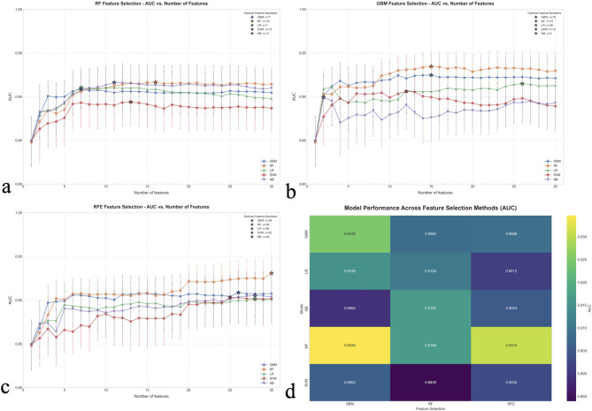
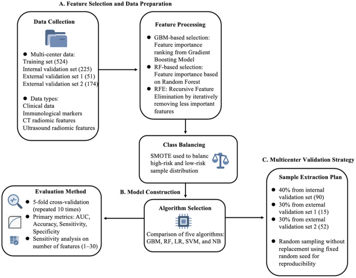
Methods: We analyzed 974 PTC patients from three medical centers in China using a multi-modal approach integrating: (1) clinical indicators, (2) immunological indices, (3) ultrasound radiomics features, and (4) CT radiomics features. Our methodology employed gradient boosting machine for feature selection and random forest for classification, with model interpretability provided through SHapley Additive exPlanations (SHAP) analysis. The model was validated on internal (n = 225) and two external cohorts (n = 51, n = 174).
Results: The final 15-feature model achieved AUCs of 0.91, 0.84, and 0.77 across validation cohorts, improving to 0.96, 0.95, and 0.89 after cohort-specific refitting. SHAP analysis revealed CT texture features, ultrasound morphological features, and immune-inflammatory markers as key predictors, with consistent patterns across validation sites despite center-specific variations. Subgroup analysis showed superior performance in tumors > 1 cm and patients without extrathyroidal extension.
Conclusion: Our multi-modal machine learning approach provides accurate preoperative risk stratification for PTC with robust cross-center applicability. This computational framework for integrating heterogeneous imaging and clinical data demonstrates the potential of multi-modal joint learning in healthcare imaging to transform clinical decision-making by enabling personalized treatment planning.
Cancer ImagingONCOLOGY-RADIOLOGY, NUCLEAR MEDICINE & MEDICAL IMAGING
CiteScore
7.00
自引率
0.00%
发文量
66
审稿时长
>12 weeks
期刊介绍:
Cancer Imaging is an open access, peer-reviewed journal publishing original articles, reviews and editorials written by expert international radiologists working in oncology.
The journal encompasses CT, MR, PET, ultrasound, radionuclide and multimodal imaging in all kinds of malignant tumours, plus new developments, techniques and innovations. Topics of interest include:
Breast Imaging
Chest
Complications of treatment
Ear, Nose & Throat
Gastrointestinal
Hepatobiliary & Pancreatic
Imaging biomarkers
Interventional
Lymphoma
Measurement of tumour response
Molecular functional imaging
Musculoskeletal
Neuro oncology
Nuclear Medicine
Paediatric.


 求助内容:
求助内容: 应助结果提醒方式:


