Somya Singhal, Sanyam K Mahajan, Sanjeev Jha, Vivek Singh, Vinita E Mani, Vimal K Paliwal
{"title":"平山病患者疾病进展的放射学和电生理学相关性。","authors":"Somya Singhal, Sanyam K Mahajan, Sanjeev Jha, Vivek Singh, Vinita E Mani, Vimal K Paliwal","doi":"10.4103/aian.aian_8_25","DOIUrl":null,"url":null,"abstract":"<p><strong>Background and objectives: </strong>To correlate the distribution of neurogenic motor unit potentials in the upper limb (s) and the extent of anterior displacement of the cervical duramater on neck flexion with the progression of weakness/atrophy in Hirayama disease.</p><p><strong>Methods: </strong>Consecutive patients with distal Hirayama disease were classified as distal group (neurogenic potential in C7-T1 innervated muscles), proximal (neurogenic potentials C5-T1 muscles), and contralateral group (neurogenic potentials in contralateral hand/arm). Based on the extent of anterior dural displacement on neck-flexed cervical magnetic resonance imaging, patients were classified as anterior dural displacement across the C5 vertebra and anterior dural displacement at C5 vertebra and below. The disease progression at 1 year was correlated with the distribution of neurogenic potentials and the extent of anterior dural displacement.</p><p><strong>Results: </strong>Twenty-eight patients (mean age, 17.41 ± 2.30 years; all males) were included. Eleven (39.2%), 17 (60.7%), and 22 (78%) patients were in proximal, distal, and contralateral groups, respectively. Twenty-three (82%) had anterior dural displacement across the C5 vertebra, whereas 5 (17%) had anterior dural displacement at C5 vertebra and below. Ipsilateral disease progression was seen in 15 (53%) and contralateral progression in 25 (89%) (new onset in 7 [25%]). No patient showed progression in shoulder/arm muscles. The proximal group had a significantly larger extent of anterior dural displacement. However, there was no correlation of disease progression with either the distribution of neurogenic motor unit potentials or the extent of cervical dural displacement on neck flexion.</p><p><strong>Conclusions: </strong>The extent of anterior dural displacement on neck flexion and neurogenic motor unit potentials in proximal, distal, or contralateral upper limb did not correlate with progression of muscle weakness/atrophy in Hirayama disease at 1 year.</p>","PeriodicalId":8036,"journal":{"name":"Annals of Indian Academy of Neurology","volume":" ","pages":"527-534"},"PeriodicalIF":1.8000,"publicationDate":"2025-07-01","publicationTypes":"Journal Article","fieldsOfStudy":null,"isOpenAccess":false,"openAccessPdf":"https://www.ncbi.nlm.nih.gov/pmc/articles/PMC12393853/pdf/","citationCount":"0","resultStr":"{\"title\":\"Radiological and Electrophysiological Correlates of Disease Progression in Patients with Hirayama Disease.\",\"authors\":\"Somya Singhal, Sanyam K Mahajan, Sanjeev Jha, Vivek Singh, Vinita E Mani, Vimal K Paliwal\",\"doi\":\"10.4103/aian.aian_8_25\",\"DOIUrl\":null,\"url\":null,\"abstract\":\"<p><strong>Background and objectives: </strong>To correlate the distribution of neurogenic motor unit potentials in the upper limb (s) and the extent of anterior displacement of the cervical duramater on neck flexion with the progression of weakness/atrophy in Hirayama disease.</p><p><strong>Methods: </strong>Consecutive patients with distal Hirayama disease were classified as distal group (neurogenic potential in C7-T1 innervated muscles), proximal (neurogenic potentials C5-T1 muscles), and contralateral group (neurogenic potentials in contralateral hand/arm). Based on the extent of anterior dural displacement on neck-flexed cervical magnetic resonance imaging, patients were classified as anterior dural displacement across the C5 vertebra and anterior dural displacement at C5 vertebra and below. The disease progression at 1 year was correlated with the distribution of neurogenic potentials and the extent of anterior dural displacement.</p><p><strong>Results: </strong>Twenty-eight patients (mean age, 17.41 ± 2.30 years; all males) were included. Eleven (39.2%), 17 (60.7%), and 22 (78%) patients were in proximal, distal, and contralateral groups, respectively. Twenty-three (82%) had anterior dural displacement across the C5 vertebra, whereas 5 (17%) had anterior dural displacement at C5 vertebra and below. Ipsilateral disease progression was seen in 15 (53%) and contralateral progression in 25 (89%) (new onset in 7 [25%]). No patient showed progression in shoulder/arm muscles. The proximal group had a significantly larger extent of anterior dural displacement. However, there was no correlation of disease progression with either the distribution of neurogenic motor unit potentials or the extent of cervical dural displacement on neck flexion.</p><p><strong>Conclusions: </strong>The extent of anterior dural displacement on neck flexion and neurogenic motor unit potentials in proximal, distal, or contralateral upper limb did not correlate with progression of muscle weakness/atrophy in Hirayama disease at 1 year.</p>\",\"PeriodicalId\":8036,\"journal\":{\"name\":\"Annals of Indian Academy of Neurology\",\"volume\":\" \",\"pages\":\"527-534\"},\"PeriodicalIF\":1.8000,\"publicationDate\":\"2025-07-01\",\"publicationTypes\":\"Journal Article\",\"fieldsOfStudy\":null,\"isOpenAccess\":false,\"openAccessPdf\":\"https://www.ncbi.nlm.nih.gov/pmc/articles/PMC12393853/pdf/\",\"citationCount\":\"0\",\"resultStr\":null,\"platform\":\"Semanticscholar\",\"paperid\":null,\"PeriodicalName\":\"Annals of Indian Academy of Neurology\",\"FirstCategoryId\":\"3\",\"ListUrlMain\":\"https://doi.org/10.4103/aian.aian_8_25\",\"RegionNum\":4,\"RegionCategory\":\"医学\",\"ArticlePicture\":[],\"TitleCN\":null,\"AbstractTextCN\":null,\"PMCID\":null,\"EPubDate\":\"2025/8/7 0:00:00\",\"PubModel\":\"Epub\",\"JCR\":\"Q3\",\"JCRName\":\"CLINICAL NEUROLOGY\",\"Score\":null,\"Total\":0}","platform":"Semanticscholar","paperid":null,"PeriodicalName":"Annals of Indian Academy of Neurology","FirstCategoryId":"3","ListUrlMain":"https://doi.org/10.4103/aian.aian_8_25","RegionNum":4,"RegionCategory":"医学","ArticlePicture":[],"TitleCN":null,"AbstractTextCN":null,"PMCID":null,"EPubDate":"2025/8/7 0:00:00","PubModel":"Epub","JCR":"Q3","JCRName":"CLINICAL NEUROLOGY","Score":null,"Total":0}
Radiological and Electrophysiological Correlates of Disease Progression in Patients with Hirayama Disease.
Background and objectives: To correlate the distribution of neurogenic motor unit potentials in the upper limb (s) and the extent of anterior displacement of the cervical duramater on neck flexion with the progression of weakness/atrophy in Hirayama disease.
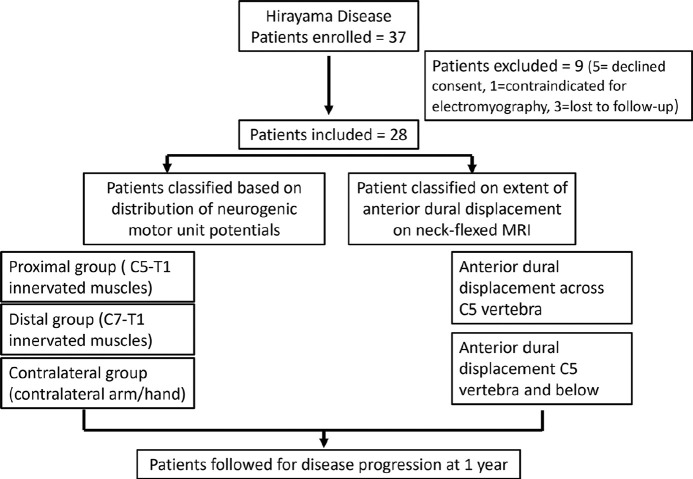
Methods: Consecutive patients with distal Hirayama disease were classified as distal group (neurogenic potential in C7-T1 innervated muscles), proximal (neurogenic potentials C5-T1 muscles), and contralateral group (neurogenic potentials in contralateral hand/arm). Based on the extent of anterior dural displacement on neck-flexed cervical magnetic resonance imaging, patients were classified as anterior dural displacement across the C5 vertebra and anterior dural displacement at C5 vertebra and below. The disease progression at 1 year was correlated with the distribution of neurogenic potentials and the extent of anterior dural displacement.
Results: Twenty-eight patients (mean age, 17.41 ± 2.30 years; all males) were included. Eleven (39.2%), 17 (60.7%), and 22 (78%) patients were in proximal, distal, and contralateral groups, respectively. Twenty-three (82%) had anterior dural displacement across the C5 vertebra, whereas 5 (17%) had anterior dural displacement at C5 vertebra and below. Ipsilateral disease progression was seen in 15 (53%) and contralateral progression in 25 (89%) (new onset in 7 [25%]). No patient showed progression in shoulder/arm muscles. The proximal group had a significantly larger extent of anterior dural displacement. However, there was no correlation of disease progression with either the distribution of neurogenic motor unit potentials or the extent of cervical dural displacement on neck flexion.
Conclusions: The extent of anterior dural displacement on neck flexion and neurogenic motor unit potentials in proximal, distal, or contralateral upper limb did not correlate with progression of muscle weakness/atrophy in Hirayama disease at 1 year.
期刊介绍:
The journal has a clinical foundation and has been utilized most by clinical neurologists for improving the practice of neurology. While the focus is on neurology in India, the journal publishes manuscripts of high value from all parts of the world. Journal publishes reviews of various types, original articles, short communications, interesting images and case reports. The journal respects the scientific submission of its authors and believes in following an expeditious double-blind peer review process and endeavors to complete the review process within scheduled time frame. A significant effort from the author and the journal perhaps enables to strike an equilibrium to meet the professional expectations of the peers in the world of scientific publication. AIAN believes in safeguarding the privacy rights of human subjects. In order to comply with it, the journal instructs all authors when uploading the manuscript to also add the ethical clearance (human/animals)/ informed consent of subject in the manuscript. This applies to the study/case report that involves animal/human subjects/human specimens e.g. extracted tooth part/soft tissue for biopsy/in vitro analysis.
 求助内容:
求助内容: 应助结果提醒方式:


