{"title":"前列腺的深度学习重建提高了t2加权成像的图像质量和获取时间。","authors":"Daichi Kobayashi, Hayato Tomita, Tsuyoshi Morimoto, Yuki Deguchi, Hirofumi Fukuchi, Hikaru Ishida, Kumie Miyakawa, Yasuyuki Kobayashi, Hidefumi Mimura","doi":"10.18999/nagjms.87.2.264","DOIUrl":null,"url":null,"abstract":"<p><p>We compared the qualitative and quantitative quality of prostate conventional T2-weighted imaging and T2-weighted imaging with deep-learning reconstruction. Patients with suspected prostate cancer undergoing magnetic resonance imaging between April 2022 and June 2023 were included. Quantitative analysis was performed to determine the signal-to-noise and contrast ratios of the perirectal fat tissue, internal obturator muscle, and pubic tubercle. Eight periprostatic anatomical structures, overall image quality, and motion artifacts were evaluated by two radiologists using 5- or 4-point scales. Qualitative analysis results were compared to determine the agreement between the two radiologists. In total, 106 patients (mean age: 71 ± 8.3 years; 106 men) were included in this study. The acquisition time for conventional T2-weighted imaging and T2-weighted imaging with deep-learning reconstruction was 4 min and 16 s and 2 min and 12 s, respectively. The signal-to-noise ratio of the perirectal fat tissue and internal obturator muscle and contrast ratio of fat/muscle and bone/muscle determined via T2-weighted imaging with deep-learning reconstruction were significantly superior to those determined via conventional T2-weighted imaging (both <i>p</i> < 0.01). Compared with conventional T2-weighted imaging, T2-weighted imaging with deep-learning reconstruction showed significant improvement in the visualization of the periprostatic anatomy, overall image quality, and motion artifacts (both <i>p</i> < 0.05). Compared with conventional methods, T2-weighted imaging with deep-learning reconstruction facilitated the acquisition of good-quality magnetic resonance images of the prostate within a shorter acquisition time. T2-weighted imaging with deep-learning reconstruction will aid clinicians in diagnosing prostate cancer with shortened acquisition time while maintaining quantitative and qualitative image properties.</p>","PeriodicalId":49014,"journal":{"name":"Nagoya Journal of Medical Science","volume":"87 2","pages":"264-271"},"PeriodicalIF":0.9000,"publicationDate":"2025-05-01","publicationTypes":"Journal Article","fieldsOfStudy":null,"isOpenAccess":false,"openAccessPdf":"https://www.ncbi.nlm.nih.gov/pmc/articles/PMC12320318/pdf/","citationCount":"0","resultStr":"{\"title\":\"Deep-learning reconstruction of the prostate improves image quality and acquisition time in T2-weighted imaging.\",\"authors\":\"Daichi Kobayashi, Hayato Tomita, Tsuyoshi Morimoto, Yuki Deguchi, Hirofumi Fukuchi, Hikaru Ishida, Kumie Miyakawa, Yasuyuki Kobayashi, Hidefumi Mimura\",\"doi\":\"10.18999/nagjms.87.2.264\",\"DOIUrl\":null,\"url\":null,\"abstract\":\"<p><p>We compared the qualitative and quantitative quality of prostate conventional T2-weighted imaging and T2-weighted imaging with deep-learning reconstruction. Patients with suspected prostate cancer undergoing magnetic resonance imaging between April 2022 and June 2023 were included. Quantitative analysis was performed to determine the signal-to-noise and contrast ratios of the perirectal fat tissue, internal obturator muscle, and pubic tubercle. Eight periprostatic anatomical structures, overall image quality, and motion artifacts were evaluated by two radiologists using 5- or 4-point scales. Qualitative analysis results were compared to determine the agreement between the two radiologists. In total, 106 patients (mean age: 71 ± 8.3 years; 106 men) were included in this study. The acquisition time for conventional T2-weighted imaging and T2-weighted imaging with deep-learning reconstruction was 4 min and 16 s and 2 min and 12 s, respectively. The signal-to-noise ratio of the perirectal fat tissue and internal obturator muscle and contrast ratio of fat/muscle and bone/muscle determined via T2-weighted imaging with deep-learning reconstruction were significantly superior to those determined via conventional T2-weighted imaging (both <i>p</i> < 0.01). Compared with conventional T2-weighted imaging, T2-weighted imaging with deep-learning reconstruction showed significant improvement in the visualization of the periprostatic anatomy, overall image quality, and motion artifacts (both <i>p</i> < 0.05). Compared with conventional methods, T2-weighted imaging with deep-learning reconstruction facilitated the acquisition of good-quality magnetic resonance images of the prostate within a shorter acquisition time. T2-weighted imaging with deep-learning reconstruction will aid clinicians in diagnosing prostate cancer with shortened acquisition time while maintaining quantitative and qualitative image properties.</p>\",\"PeriodicalId\":49014,\"journal\":{\"name\":\"Nagoya Journal of Medical Science\",\"volume\":\"87 2\",\"pages\":\"264-271\"},\"PeriodicalIF\":0.9000,\"publicationDate\":\"2025-05-01\",\"publicationTypes\":\"Journal Article\",\"fieldsOfStudy\":null,\"isOpenAccess\":false,\"openAccessPdf\":\"https://www.ncbi.nlm.nih.gov/pmc/articles/PMC12320318/pdf/\",\"citationCount\":\"0\",\"resultStr\":null,\"platform\":\"Semanticscholar\",\"paperid\":null,\"PeriodicalName\":\"Nagoya Journal of Medical Science\",\"FirstCategoryId\":\"3\",\"ListUrlMain\":\"https://doi.org/10.18999/nagjms.87.2.264\",\"RegionNum\":4,\"RegionCategory\":\"医学\",\"ArticlePicture\":[],\"TitleCN\":null,\"AbstractTextCN\":null,\"PMCID\":null,\"EPubDate\":\"\",\"PubModel\":\"\",\"JCR\":\"Q4\",\"JCRName\":\"MEDICINE, RESEARCH & EXPERIMENTAL\",\"Score\":null,\"Total\":0}","platform":"Semanticscholar","paperid":null,"PeriodicalName":"Nagoya Journal of Medical Science","FirstCategoryId":"3","ListUrlMain":"https://doi.org/10.18999/nagjms.87.2.264","RegionNum":4,"RegionCategory":"医学","ArticlePicture":[],"TitleCN":null,"AbstractTextCN":null,"PMCID":null,"EPubDate":"","PubModel":"","JCR":"Q4","JCRName":"MEDICINE, RESEARCH & EXPERIMENTAL","Score":null,"Total":0}
引用次数: 0
摘要
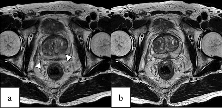
我们比较了前列腺常规t2加权成像和深度学习重建的t2加权成像的定性和定量质量。研究纳入了2022年4月至2023年6月期间接受磁共振成像的疑似前列腺癌患者。定量分析直肠周围脂肪组织、内闭孔肌和耻骨结节的信噪比和对比。8个前列腺周围解剖结构、整体图像质量和运动伪影由两名放射科医生用5或4分制进行评估。对定性分析结果进行比较,以确定两名放射科医生的一致意见。106例患者(平均年龄71±8.3岁;106名男性)纳入本研究。常规t2加权成像和深度学习重建t2加权成像的采集时间分别为4 min 16 s和2 min 12 s。深度学习重建t2加权成像直肠周围脂肪组织和内闭孔肌的信噪比、脂肪/肌肉和骨/肌肉的对比明显优于常规t2加权成像(p < 0.01)。与传统的t2加权成像相比,深度学习重建的t2加权成像在前列腺周围解剖的可视化、整体图像质量和运动伪影方面均有显著改善(p < 0.05)。与传统方法相比,深度学习重建的t2加权成像可以在更短的采集时间内获得高质量的前列腺磁共振图像。深度学习重建的t2加权成像将在保持定量和定性图像特性的同时,缩短采集时间,帮助临床医生诊断前列腺癌。
Deep-learning reconstruction of the prostate improves image quality and acquisition time in T2-weighted imaging.
We compared the qualitative and quantitative quality of prostate conventional T2-weighted imaging and T2-weighted imaging with deep-learning reconstruction. Patients with suspected prostate cancer undergoing magnetic resonance imaging between April 2022 and June 2023 were included. Quantitative analysis was performed to determine the signal-to-noise and contrast ratios of the perirectal fat tissue, internal obturator muscle, and pubic tubercle. Eight periprostatic anatomical structures, overall image quality, and motion artifacts were evaluated by two radiologists using 5- or 4-point scales. Qualitative analysis results were compared to determine the agreement between the two radiologists. In total, 106 patients (mean age: 71 ± 8.3 years; 106 men) were included in this study. The acquisition time for conventional T2-weighted imaging and T2-weighted imaging with deep-learning reconstruction was 4 min and 16 s and 2 min and 12 s, respectively. The signal-to-noise ratio of the perirectal fat tissue and internal obturator muscle and contrast ratio of fat/muscle and bone/muscle determined via T2-weighted imaging with deep-learning reconstruction were significantly superior to those determined via conventional T2-weighted imaging (both p < 0.01). Compared with conventional T2-weighted imaging, T2-weighted imaging with deep-learning reconstruction showed significant improvement in the visualization of the periprostatic anatomy, overall image quality, and motion artifacts (both p < 0.05). Compared with conventional methods, T2-weighted imaging with deep-learning reconstruction facilitated the acquisition of good-quality magnetic resonance images of the prostate within a shorter acquisition time. T2-weighted imaging with deep-learning reconstruction will aid clinicians in diagnosing prostate cancer with shortened acquisition time while maintaining quantitative and qualitative image properties.
期刊介绍:
The Journal publishes original papers in the areas of medical science and its related fields. Reviews, symposium reports, short communications, notes, case reports, hypothesis papers, medical image at a glance, video and announcements are also accepted.
Manuscripts should be in English. It is recommended that an English check of the manuscript by a competent and knowledgeable native speaker be completed before submission.

 求助内容:
求助内容: 应助结果提醒方式:


