Stefano Espenica, Edward Bellamy, Shauna Hilton, Carlo Bianco, Gail Leeming, Hanna Procter, Ferran Valls Sanchez
{"title":"猫全身反应性血管内皮瘤病伴高钙血症。","authors":"Stefano Espenica, Edward Bellamy, Shauna Hilton, Carlo Bianco, Gail Leeming, Hanna Procter, Ferran Valls Sanchez","doi":"10.1177/20551169251347078","DOIUrl":null,"url":null,"abstract":"<p><strong>Case summary: </strong>A 3-year 10-month-old spayed female domestic shorthair cat was presented for subacute progressive hyporexia, vomiting and lethargy. On presentation, the cat was dyspnoeic, and venous blood gas analysis revealed metabolic acidosis, hypercalcaemia (both total and ionised), hyperlactaemia and hyperglycaemia. Physical examination identified a 2 × 3 cm crusted cutaneous lesion on the cranium, reduced mentation, mild tachycardia, harsh bronchovesicular sounds and approximately 5% dehydration. Owing to welfare concerns, the owners elected euthanasia. Post-mortem examination revealed moderate autolytic changes. The organs most affected by vascular lesions included the heart, brain, kidneys, liver and pancreas. Histopathology revealed mild to severe multifocal intraluminal and mural proliferations of atypical endothelial cells, accompanied by multifocal thrombosis and mild perivascular oedema. Immunohistochemistry showed that the proliferating cells were negative for alpha-smooth muscle actin, and quantitative PCR for <i>Bartonella</i> species was also negative. However, 50% of the proliferating cells were positive for factor VIII. These findings supported a diagnosis of feline systemic reactive angioendotheliomatosis.</p><p><strong>Relevance and novel information: </strong>In the authors' opinion, this case contributes to the growing body of literature on this rare condition and raises the possibility of an association with hypercalcaemia.</p>","PeriodicalId":36588,"journal":{"name":"Journal of Feline Medicine and Surgery Open Reports","volume":"11 2","pages":"20551169251347078"},"PeriodicalIF":0.7000,"publicationDate":"2025-08-04","publicationTypes":"Journal Article","fieldsOfStudy":null,"isOpenAccess":false,"openAccessPdf":"https://www.ncbi.nlm.nih.gov/pmc/articles/PMC12322373/pdf/","citationCount":"0","resultStr":"{\"title\":\"Feline systemic reactive angioendotheliomatosis with hypercalcaemia.\",\"authors\":\"Stefano Espenica, Edward Bellamy, Shauna Hilton, Carlo Bianco, Gail Leeming, Hanna Procter, Ferran Valls Sanchez\",\"doi\":\"10.1177/20551169251347078\",\"DOIUrl\":null,\"url\":null,\"abstract\":\"<p><strong>Case summary: </strong>A 3-year 10-month-old spayed female domestic shorthair cat was presented for subacute progressive hyporexia, vomiting and lethargy. On presentation, the cat was dyspnoeic, and venous blood gas analysis revealed metabolic acidosis, hypercalcaemia (both total and ionised), hyperlactaemia and hyperglycaemia. Physical examination identified a 2 × 3 cm crusted cutaneous lesion on the cranium, reduced mentation, mild tachycardia, harsh bronchovesicular sounds and approximately 5% dehydration. Owing to welfare concerns, the owners elected euthanasia. Post-mortem examination revealed moderate autolytic changes. The organs most affected by vascular lesions included the heart, brain, kidneys, liver and pancreas. Histopathology revealed mild to severe multifocal intraluminal and mural proliferations of atypical endothelial cells, accompanied by multifocal thrombosis and mild perivascular oedema. Immunohistochemistry showed that the proliferating cells were negative for alpha-smooth muscle actin, and quantitative PCR for <i>Bartonella</i> species was also negative. However, 50% of the proliferating cells were positive for factor VIII. These findings supported a diagnosis of feline systemic reactive angioendotheliomatosis.</p><p><strong>Relevance and novel information: </strong>In the authors' opinion, this case contributes to the growing body of literature on this rare condition and raises the possibility of an association with hypercalcaemia.</p>\",\"PeriodicalId\":36588,\"journal\":{\"name\":\"Journal of Feline Medicine and Surgery Open Reports\",\"volume\":\"11 2\",\"pages\":\"20551169251347078\"},\"PeriodicalIF\":0.7000,\"publicationDate\":\"2025-08-04\",\"publicationTypes\":\"Journal Article\",\"fieldsOfStudy\":null,\"isOpenAccess\":false,\"openAccessPdf\":\"https://www.ncbi.nlm.nih.gov/pmc/articles/PMC12322373/pdf/\",\"citationCount\":\"0\",\"resultStr\":null,\"platform\":\"Semanticscholar\",\"paperid\":null,\"PeriodicalName\":\"Journal of Feline Medicine and Surgery Open Reports\",\"FirstCategoryId\":\"1085\",\"ListUrlMain\":\"https://doi.org/10.1177/20551169251347078\",\"RegionNum\":0,\"RegionCategory\":null,\"ArticlePicture\":[],\"TitleCN\":null,\"AbstractTextCN\":null,\"PMCID\":null,\"EPubDate\":\"2025/7/1 0:00:00\",\"PubModel\":\"eCollection\",\"JCR\":\"Q3\",\"JCRName\":\"VETERINARY SCIENCES\",\"Score\":null,\"Total\":0}","platform":"Semanticscholar","paperid":null,"PeriodicalName":"Journal of Feline Medicine and Surgery Open Reports","FirstCategoryId":"1085","ListUrlMain":"https://doi.org/10.1177/20551169251347078","RegionNum":0,"RegionCategory":null,"ArticlePicture":[],"TitleCN":null,"AbstractTextCN":null,"PMCID":null,"EPubDate":"2025/7/1 0:00:00","PubModel":"eCollection","JCR":"Q3","JCRName":"VETERINARY SCIENCES","Score":null,"Total":0}
Feline systemic reactive angioendotheliomatosis with hypercalcaemia.
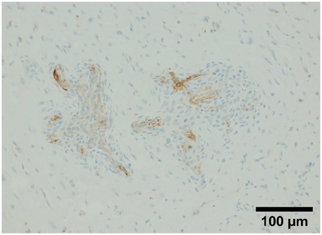
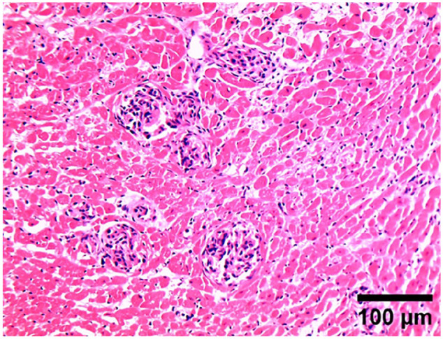
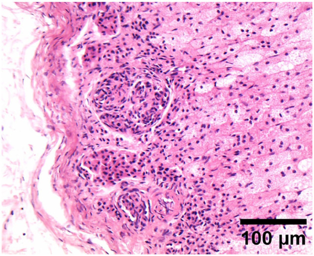
Case summary: A 3-year 10-month-old spayed female domestic shorthair cat was presented for subacute progressive hyporexia, vomiting and lethargy. On presentation, the cat was dyspnoeic, and venous blood gas analysis revealed metabolic acidosis, hypercalcaemia (both total and ionised), hyperlactaemia and hyperglycaemia. Physical examination identified a 2 × 3 cm crusted cutaneous lesion on the cranium, reduced mentation, mild tachycardia, harsh bronchovesicular sounds and approximately 5% dehydration. Owing to welfare concerns, the owners elected euthanasia. Post-mortem examination revealed moderate autolytic changes. The organs most affected by vascular lesions included the heart, brain, kidneys, liver and pancreas. Histopathology revealed mild to severe multifocal intraluminal and mural proliferations of atypical endothelial cells, accompanied by multifocal thrombosis and mild perivascular oedema. Immunohistochemistry showed that the proliferating cells were negative for alpha-smooth muscle actin, and quantitative PCR for Bartonella species was also negative. However, 50% of the proliferating cells were positive for factor VIII. These findings supported a diagnosis of feline systemic reactive angioendotheliomatosis.
Relevance and novel information: In the authors' opinion, this case contributes to the growing body of literature on this rare condition and raises the possibility of an association with hypercalcaemia.

 求助内容:
求助内容: 应助结果提醒方式:


