{"title":"腹股沟肿瘤切除后外阴肌上皮瘤样肿瘤1例。","authors":"Kei Urakami, Hiroaki Saito, Akimitsu Tanio, Yoichiro Tada, Yoshinori Yamada, Yutaka Yamashiro, Yumi Yamaguchi","doi":"10.70352/scrj.cr.25-0237","DOIUrl":null,"url":null,"abstract":"<p><strong>Introduction: </strong>Myoepithelioma-like tumor of the vulvar region (MELTVR) is a mesenchymal neoplasm first reported in 2015 and typically develops from the inguinal to the vulvar regions of adult women.</p><p><strong>Case presentation: </strong>Here we report the case of a 42-year-old woman who presented with right inguinal tumor. The tumor had recently increased in size continuously. Computed tomography (CT) showed a homogeneous neoplastic lesion along the uterine cord in the right inguinal region and marginal resection was performed. Pathological examination revealed a well-defined tumor. And there were areas of epithelial-like tumor cells arranged in a reticular or cord-like pattern against a background of mucinous stroma, and areas of spindle-shaped cells growing in mucinous substrate with transition from epithelial cells. The nucleus was irregular in size and shape. Necrotic nests were scattered in the tumor. Immunohistological examination showed that the tumor cells were positive for epithelial membrane antigen (EMA), estrogen receptor (ER), and progesterone receptor (PgR). Alpha-smooth muscle actin (α-SMA) was slightly positive. The tumor was negative for cytokeratin AE1/AE3, p63, desmin, CD34, S100, glial fibrillary acidic protein (GFAP), and SOX10. Loss of INI1 protein expression was also confirmed. The patient was suspected of having high-grade myoepithelioma on pathological diagnosis at our hospital. However, immunohistological findings led to the diagnosis of MELTVR. The patient underwent additional wide excision and has been alive 10 months postoperatively without recurrence.</p><p><strong>Conclusions: </strong>Due to its rarity, it is difficult to make preoperative diagnosis of MELTVR. Awareness of this condition can contribute to accurate diagnosis and appropriate management in adult female patients presenting with swelling extending from the inguinal to the vulvar regions.</p>","PeriodicalId":22096,"journal":{"name":"Surgical Case Reports","volume":"11 1","pages":""},"PeriodicalIF":0.7000,"publicationDate":"2025-01-01","publicationTypes":"Journal Article","fieldsOfStudy":null,"isOpenAccess":false,"openAccessPdf":"https://www.ncbi.nlm.nih.gov/pmc/articles/PMC12324924/pdf/","citationCount":"0","resultStr":"{\"title\":\"A Case of Myoepithelioma-Like Tumor of the Vulvar Region after Removal of an Inguinal Tumor.\",\"authors\":\"Kei Urakami, Hiroaki Saito, Akimitsu Tanio, Yoichiro Tada, Yoshinori Yamada, Yutaka Yamashiro, Yumi Yamaguchi\",\"doi\":\"10.70352/scrj.cr.25-0237\",\"DOIUrl\":null,\"url\":null,\"abstract\":\"<p><strong>Introduction: </strong>Myoepithelioma-like tumor of the vulvar region (MELTVR) is a mesenchymal neoplasm first reported in 2015 and typically develops from the inguinal to the vulvar regions of adult women.</p><p><strong>Case presentation: </strong>Here we report the case of a 42-year-old woman who presented with right inguinal tumor. The tumor had recently increased in size continuously. Computed tomography (CT) showed a homogeneous neoplastic lesion along the uterine cord in the right inguinal region and marginal resection was performed. Pathological examination revealed a well-defined tumor. And there were areas of epithelial-like tumor cells arranged in a reticular or cord-like pattern against a background of mucinous stroma, and areas of spindle-shaped cells growing in mucinous substrate with transition from epithelial cells. The nucleus was irregular in size and shape. Necrotic nests were scattered in the tumor. Immunohistological examination showed that the tumor cells were positive for epithelial membrane antigen (EMA), estrogen receptor (ER), and progesterone receptor (PgR). Alpha-smooth muscle actin (α-SMA) was slightly positive. The tumor was negative for cytokeratin AE1/AE3, p63, desmin, CD34, S100, glial fibrillary acidic protein (GFAP), and SOX10. Loss of INI1 protein expression was also confirmed. The patient was suspected of having high-grade myoepithelioma on pathological diagnosis at our hospital. However, immunohistological findings led to the diagnosis of MELTVR. The patient underwent additional wide excision and has been alive 10 months postoperatively without recurrence.</p><p><strong>Conclusions: </strong>Due to its rarity, it is difficult to make preoperative diagnosis of MELTVR. Awareness of this condition can contribute to accurate diagnosis and appropriate management in adult female patients presenting with swelling extending from the inguinal to the vulvar regions.</p>\",\"PeriodicalId\":22096,\"journal\":{\"name\":\"Surgical Case Reports\",\"volume\":\"11 1\",\"pages\":\"\"},\"PeriodicalIF\":0.7000,\"publicationDate\":\"2025-01-01\",\"publicationTypes\":\"Journal Article\",\"fieldsOfStudy\":null,\"isOpenAccess\":false,\"openAccessPdf\":\"https://www.ncbi.nlm.nih.gov/pmc/articles/PMC12324924/pdf/\",\"citationCount\":\"0\",\"resultStr\":null,\"platform\":\"Semanticscholar\",\"paperid\":null,\"PeriodicalName\":\"Surgical Case Reports\",\"FirstCategoryId\":\"1085\",\"ListUrlMain\":\"https://doi.org/10.70352/scrj.cr.25-0237\",\"RegionNum\":0,\"RegionCategory\":null,\"ArticlePicture\":[],\"TitleCN\":null,\"AbstractTextCN\":null,\"PMCID\":null,\"EPubDate\":\"2025/8/5 0:00:00\",\"PubModel\":\"Epub\",\"JCR\":\"Q4\",\"JCRName\":\"SURGERY\",\"Score\":null,\"Total\":0}","platform":"Semanticscholar","paperid":null,"PeriodicalName":"Surgical Case Reports","FirstCategoryId":"1085","ListUrlMain":"https://doi.org/10.70352/scrj.cr.25-0237","RegionNum":0,"RegionCategory":null,"ArticlePicture":[],"TitleCN":null,"AbstractTextCN":null,"PMCID":null,"EPubDate":"2025/8/5 0:00:00","PubModel":"Epub","JCR":"Q4","JCRName":"SURGERY","Score":null,"Total":0}
A Case of Myoepithelioma-Like Tumor of the Vulvar Region after Removal of an Inguinal Tumor.
Introduction: Myoepithelioma-like tumor of the vulvar region (MELTVR) is a mesenchymal neoplasm first reported in 2015 and typically develops from the inguinal to the vulvar regions of adult women.
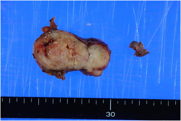
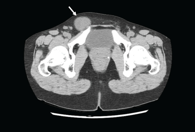
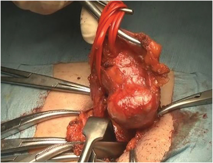
Case presentation: Here we report the case of a 42-year-old woman who presented with right inguinal tumor. The tumor had recently increased in size continuously. Computed tomography (CT) showed a homogeneous neoplastic lesion along the uterine cord in the right inguinal region and marginal resection was performed. Pathological examination revealed a well-defined tumor. And there were areas of epithelial-like tumor cells arranged in a reticular or cord-like pattern against a background of mucinous stroma, and areas of spindle-shaped cells growing in mucinous substrate with transition from epithelial cells. The nucleus was irregular in size and shape. Necrotic nests were scattered in the tumor. Immunohistological examination showed that the tumor cells were positive for epithelial membrane antigen (EMA), estrogen receptor (ER), and progesterone receptor (PgR). Alpha-smooth muscle actin (α-SMA) was slightly positive. The tumor was negative for cytokeratin AE1/AE3, p63, desmin, CD34, S100, glial fibrillary acidic protein (GFAP), and SOX10. Loss of INI1 protein expression was also confirmed. The patient was suspected of having high-grade myoepithelioma on pathological diagnosis at our hospital. However, immunohistological findings led to the diagnosis of MELTVR. The patient underwent additional wide excision and has been alive 10 months postoperatively without recurrence.
Conclusions: Due to its rarity, it is difficult to make preoperative diagnosis of MELTVR. Awareness of this condition can contribute to accurate diagnosis and appropriate management in adult female patients presenting with swelling extending from the inguinal to the vulvar regions.

 求助内容:
求助内容: 应助结果提醒方式:


