{"title":"乳突旁体独特有丝分裂机制的发现。","authors":"Bungo Akiyoshi, Drahomíra Faktorová, Julius Lukeš","doi":"10.1098/rsob.250096","DOIUrl":null,"url":null,"abstract":"<p><p>Diplonemids are highly diverse and abundant marine plankton with significant ecological importance. However, little is known about their biology, even in the model diplonemid <i>Paradiplonema papillatum</i> whose genome sequence is available. Examining the subcellular localization of proteins using fluorescence microscopy is a powerful approach to infer their putative function. Here, we report a plasmid-based method that enables YFP-tagging of a gene at the endogenous locus. By examining the localization of proteins whose homologs are involved in chromosome organization or segregation in other eukaryotes, we discovered several notable features in mitotically dividing <i>P. papillatum</i> cells. Cohesin is enriched on condensed interphase chromatin. During mitosis, chromosomes organize into two rings (termed mitotic rings herein) that surround the elongating nucleolus and align on a bipolar spindle. Homologs of chromosomal passenger complex components (INCENP, two Aurora kinases and KIN-A), a CLK1 kinase, meiotic chromosome axis protein SYCP2L1, spindle checkpoint protein Mad1 and microtubule regulator XMAP215 localize in between the two mitotic rings. In contrast, a Mad2 homolog localizes near basal bodies as in trypanosomes. By representing the first molecular characterization of mitotic mechanisms in <i>P. papillatum</i> and raising many questions, this study forms the foundation for dissecting mitotic mechanisms in diplonemids.</p>","PeriodicalId":19629,"journal":{"name":"Open Biology","volume":"15 8","pages":"250096"},"PeriodicalIF":3.6000,"publicationDate":"2025-08-01","publicationTypes":"Journal Article","fieldsOfStudy":null,"isOpenAccess":false,"openAccessPdf":"https://www.ncbi.nlm.nih.gov/pmc/articles/PMC12324889/pdf/","citationCount":"0","resultStr":"{\"title\":\"Discovery of unique mitotic mechanisms in <i>Paradiplonema papillatum</i>.\",\"authors\":\"Bungo Akiyoshi, Drahomíra Faktorová, Julius Lukeš\",\"doi\":\"10.1098/rsob.250096\",\"DOIUrl\":null,\"url\":null,\"abstract\":\"<p><p>Diplonemids are highly diverse and abundant marine plankton with significant ecological importance. However, little is known about their biology, even in the model diplonemid <i>Paradiplonema papillatum</i> whose genome sequence is available. Examining the subcellular localization of proteins using fluorescence microscopy is a powerful approach to infer their putative function. Here, we report a plasmid-based method that enables YFP-tagging of a gene at the endogenous locus. By examining the localization of proteins whose homologs are involved in chromosome organization or segregation in other eukaryotes, we discovered several notable features in mitotically dividing <i>P. papillatum</i> cells. Cohesin is enriched on condensed interphase chromatin. During mitosis, chromosomes organize into two rings (termed mitotic rings herein) that surround the elongating nucleolus and align on a bipolar spindle. Homologs of chromosomal passenger complex components (INCENP, two Aurora kinases and KIN-A), a CLK1 kinase, meiotic chromosome axis protein SYCP2L1, spindle checkpoint protein Mad1 and microtubule regulator XMAP215 localize in between the two mitotic rings. In contrast, a Mad2 homolog localizes near basal bodies as in trypanosomes. By representing the first molecular characterization of mitotic mechanisms in <i>P. papillatum</i> and raising many questions, this study forms the foundation for dissecting mitotic mechanisms in diplonemids.</p>\",\"PeriodicalId\":19629,\"journal\":{\"name\":\"Open Biology\",\"volume\":\"15 8\",\"pages\":\"250096\"},\"PeriodicalIF\":3.6000,\"publicationDate\":\"2025-08-01\",\"publicationTypes\":\"Journal Article\",\"fieldsOfStudy\":null,\"isOpenAccess\":false,\"openAccessPdf\":\"https://www.ncbi.nlm.nih.gov/pmc/articles/PMC12324889/pdf/\",\"citationCount\":\"0\",\"resultStr\":null,\"platform\":\"Semanticscholar\",\"paperid\":null,\"PeriodicalName\":\"Open Biology\",\"FirstCategoryId\":\"99\",\"ListUrlMain\":\"https://doi.org/10.1098/rsob.250096\",\"RegionNum\":3,\"RegionCategory\":\"生物学\",\"ArticlePicture\":[],\"TitleCN\":null,\"AbstractTextCN\":null,\"PMCID\":null,\"EPubDate\":\"2025/8/6 0:00:00\",\"PubModel\":\"Epub\",\"JCR\":\"Q1\",\"JCRName\":\"BIOCHEMISTRY & MOLECULAR BIOLOGY\",\"Score\":null,\"Total\":0}","platform":"Semanticscholar","paperid":null,"PeriodicalName":"Open Biology","FirstCategoryId":"99","ListUrlMain":"https://doi.org/10.1098/rsob.250096","RegionNum":3,"RegionCategory":"生物学","ArticlePicture":[],"TitleCN":null,"AbstractTextCN":null,"PMCID":null,"EPubDate":"2025/8/6 0:00:00","PubModel":"Epub","JCR":"Q1","JCRName":"BIOCHEMISTRY & MOLECULAR BIOLOGY","Score":null,"Total":0}
Discovery of unique mitotic mechanisms in Paradiplonema papillatum.
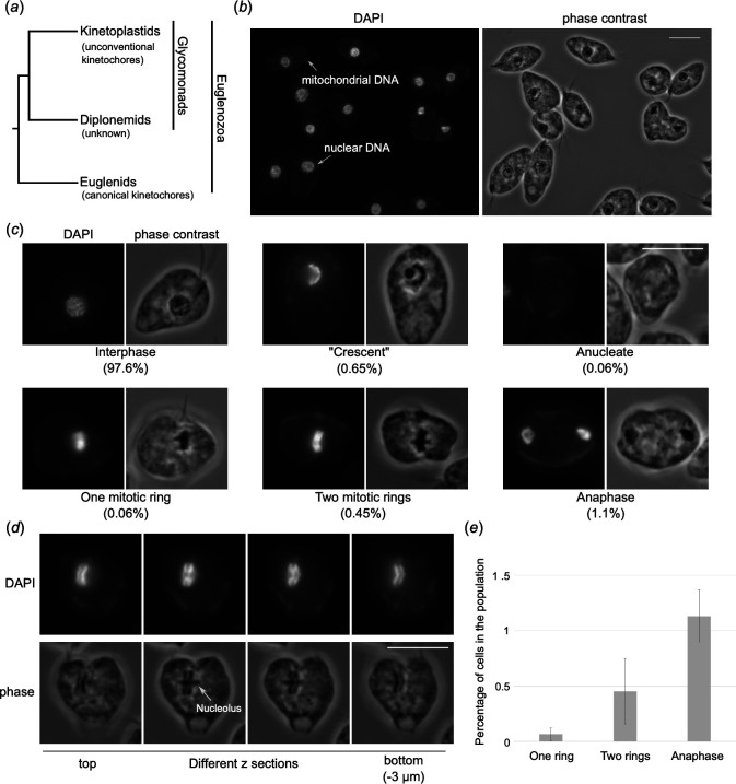
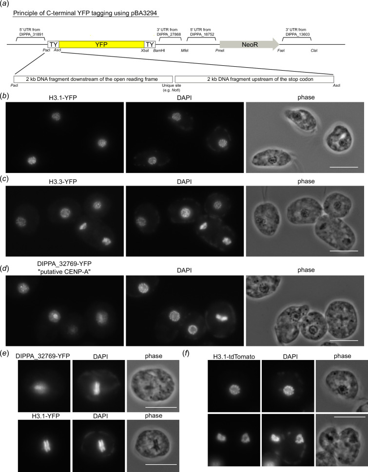
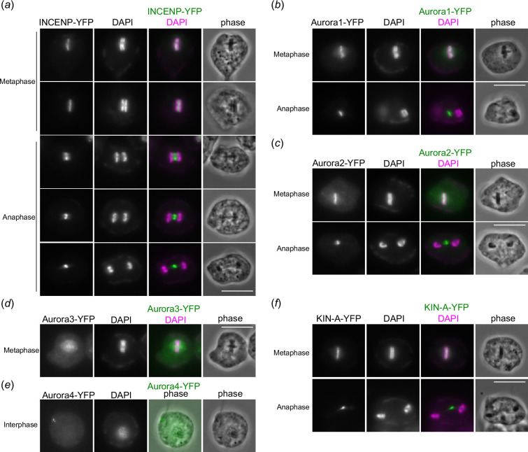
Diplonemids are highly diverse and abundant marine plankton with significant ecological importance. However, little is known about their biology, even in the model diplonemid Paradiplonema papillatum whose genome sequence is available. Examining the subcellular localization of proteins using fluorescence microscopy is a powerful approach to infer their putative function. Here, we report a plasmid-based method that enables YFP-tagging of a gene at the endogenous locus. By examining the localization of proteins whose homologs are involved in chromosome organization or segregation in other eukaryotes, we discovered several notable features in mitotically dividing P. papillatum cells. Cohesin is enriched on condensed interphase chromatin. During mitosis, chromosomes organize into two rings (termed mitotic rings herein) that surround the elongating nucleolus and align on a bipolar spindle. Homologs of chromosomal passenger complex components (INCENP, two Aurora kinases and KIN-A), a CLK1 kinase, meiotic chromosome axis protein SYCP2L1, spindle checkpoint protein Mad1 and microtubule regulator XMAP215 localize in between the two mitotic rings. In contrast, a Mad2 homolog localizes near basal bodies as in trypanosomes. By representing the first molecular characterization of mitotic mechanisms in P. papillatum and raising many questions, this study forms the foundation for dissecting mitotic mechanisms in diplonemids.
期刊介绍:
Open Biology is an online journal that welcomes original, high impact research in cell and developmental biology, molecular and structural biology, biochemistry, neuroscience, immunology, microbiology and genetics.


 求助内容:
求助内容: 应助结果提醒方式:


