{"title":"鉴别脐膨出与假性脐膨出,突出超声检查缺陷1例。","authors":"Prajwal Dahal, Rudra Prasad Upadhyaya, Ongden Yonjen Tamang, Sabina Parajuli","doi":"10.1186/s13256-025-05449-y","DOIUrl":null,"url":null,"abstract":"<p><strong>Background: </strong>Omphalocele is a congenital anomaly where abdominal contents herniate through a defect in the fetal abdominal wall, covered by peritoneum and amnion. It is associated with high mortality and other anomalies. Pseudo-omphalocele is a potential pitfall in antenatal ultrasonography, where a transient bulge of abdominal contents may appear owing to factors such as a contracted uterus, placenta, or excessive transducer pressure.</p><p><strong>Case report: </strong>This report presents two cases: one of true omphalocele in a twin pregnancy and another of pseudo-omphalocele, underscoring the importance of careful assessment. The first case involves a twin pregnancy at 12 weeks' gestation, conceived through assisted reproductive technique in a 38-year-old Nepali woman of Indo-Aryan ethnicity. During a routine check-up, one twin was diagnosed with omphalocele. Trans-abdominal fetal reduction of the anomalous twin was performed. The other twin progressed to term and was delivered via cesarean section at 39 weeks. The second case involved pseudo-omphalocele, observed at 15 weeks' 3 days of gestation in a 32-year-old Nepali woman of Tibeto-Burmese ethnicity. Initially, the fetal abdomen appeared to herniate, mimicking omphalocele. However, a repeat examination after 30 min showed no herniation or defect. Retrospective analysis revealed that the misdiagnosis occurred because the fetal abdomen was compressed between the contracted myometrium and placenta.</p><p><strong>Conclusion: </strong>Accurate diagnosis of omphalocele is crucial to prevent unnecessary abortions and potential professional repercussions. We recommend repeat examination after 30 min in all cases of omphalocele to prevent misdiagnosis.</p>","PeriodicalId":16236,"journal":{"name":"Journal of Medical Case Reports","volume":"19 1","pages":"388"},"PeriodicalIF":0.8000,"publicationDate":"2025-08-05","publicationTypes":"Journal Article","fieldsOfStudy":null,"isOpenAccess":false,"openAccessPdf":"https://www.ncbi.nlm.nih.gov/pmc/articles/PMC12323075/pdf/","citationCount":"0","resultStr":"{\"title\":\"Distinguishing omphalocele from pseudo-omphalocele, highlighting the ultrasonography pitfall: a case report.\",\"authors\":\"Prajwal Dahal, Rudra Prasad Upadhyaya, Ongden Yonjen Tamang, Sabina Parajuli\",\"doi\":\"10.1186/s13256-025-05449-y\",\"DOIUrl\":null,\"url\":null,\"abstract\":\"<p><strong>Background: </strong>Omphalocele is a congenital anomaly where abdominal contents herniate through a defect in the fetal abdominal wall, covered by peritoneum and amnion. It is associated with high mortality and other anomalies. Pseudo-omphalocele is a potential pitfall in antenatal ultrasonography, where a transient bulge of abdominal contents may appear owing to factors such as a contracted uterus, placenta, or excessive transducer pressure.</p><p><strong>Case report: </strong>This report presents two cases: one of true omphalocele in a twin pregnancy and another of pseudo-omphalocele, underscoring the importance of careful assessment. The first case involves a twin pregnancy at 12 weeks' gestation, conceived through assisted reproductive technique in a 38-year-old Nepali woman of Indo-Aryan ethnicity. During a routine check-up, one twin was diagnosed with omphalocele. Trans-abdominal fetal reduction of the anomalous twin was performed. The other twin progressed to term and was delivered via cesarean section at 39 weeks. The second case involved pseudo-omphalocele, observed at 15 weeks' 3 days of gestation in a 32-year-old Nepali woman of Tibeto-Burmese ethnicity. Initially, the fetal abdomen appeared to herniate, mimicking omphalocele. However, a repeat examination after 30 min showed no herniation or defect. Retrospective analysis revealed that the misdiagnosis occurred because the fetal abdomen was compressed between the contracted myometrium and placenta.</p><p><strong>Conclusion: </strong>Accurate diagnosis of omphalocele is crucial to prevent unnecessary abortions and potential professional repercussions. We recommend repeat examination after 30 min in all cases of omphalocele to prevent misdiagnosis.</p>\",\"PeriodicalId\":16236,\"journal\":{\"name\":\"Journal of Medical Case Reports\",\"volume\":\"19 1\",\"pages\":\"388\"},\"PeriodicalIF\":0.8000,\"publicationDate\":\"2025-08-05\",\"publicationTypes\":\"Journal Article\",\"fieldsOfStudy\":null,\"isOpenAccess\":false,\"openAccessPdf\":\"https://www.ncbi.nlm.nih.gov/pmc/articles/PMC12323075/pdf/\",\"citationCount\":\"0\",\"resultStr\":null,\"platform\":\"Semanticscholar\",\"paperid\":null,\"PeriodicalName\":\"Journal of Medical Case Reports\",\"FirstCategoryId\":\"1085\",\"ListUrlMain\":\"https://doi.org/10.1186/s13256-025-05449-y\",\"RegionNum\":0,\"RegionCategory\":null,\"ArticlePicture\":[],\"TitleCN\":null,\"AbstractTextCN\":null,\"PMCID\":null,\"EPubDate\":\"\",\"PubModel\":\"\",\"JCR\":\"Q3\",\"JCRName\":\"MEDICINE, GENERAL & INTERNAL\",\"Score\":null,\"Total\":0}","platform":"Semanticscholar","paperid":null,"PeriodicalName":"Journal of Medical Case Reports","FirstCategoryId":"1085","ListUrlMain":"https://doi.org/10.1186/s13256-025-05449-y","RegionNum":0,"RegionCategory":null,"ArticlePicture":[],"TitleCN":null,"AbstractTextCN":null,"PMCID":null,"EPubDate":"","PubModel":"","JCR":"Q3","JCRName":"MEDICINE, GENERAL & INTERNAL","Score":null,"Total":0}
Distinguishing omphalocele from pseudo-omphalocele, highlighting the ultrasonography pitfall: a case report.
Background: Omphalocele is a congenital anomaly where abdominal contents herniate through a defect in the fetal abdominal wall, covered by peritoneum and amnion. It is associated with high mortality and other anomalies. Pseudo-omphalocele is a potential pitfall in antenatal ultrasonography, where a transient bulge of abdominal contents may appear owing to factors such as a contracted uterus, placenta, or excessive transducer pressure.
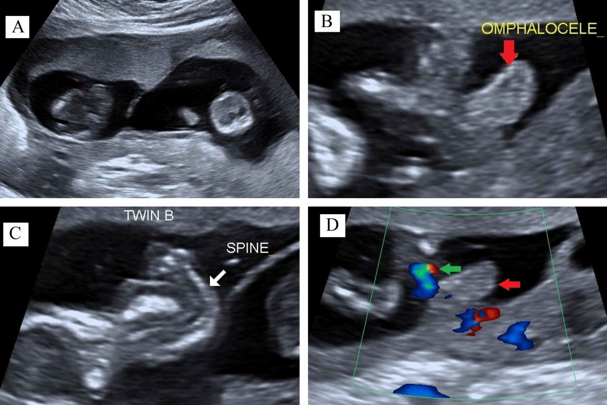
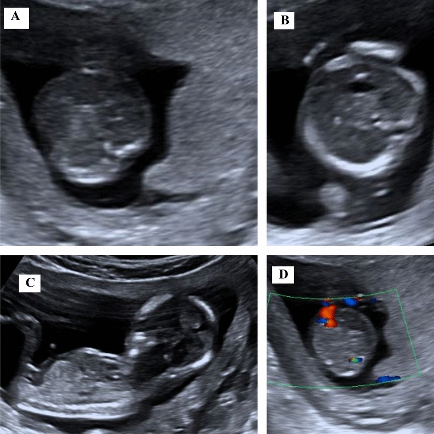
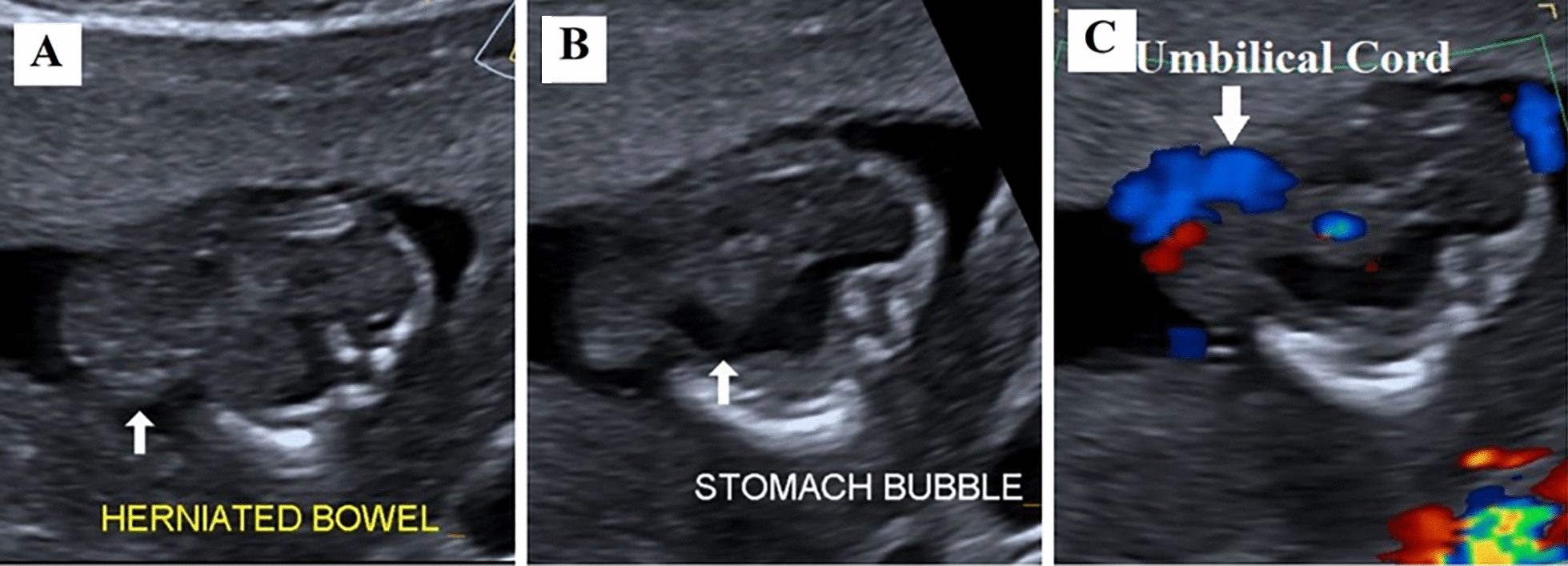
Case report: This report presents two cases: one of true omphalocele in a twin pregnancy and another of pseudo-omphalocele, underscoring the importance of careful assessment. The first case involves a twin pregnancy at 12 weeks' gestation, conceived through assisted reproductive technique in a 38-year-old Nepali woman of Indo-Aryan ethnicity. During a routine check-up, one twin was diagnosed with omphalocele. Trans-abdominal fetal reduction of the anomalous twin was performed. The other twin progressed to term and was delivered via cesarean section at 39 weeks. The second case involved pseudo-omphalocele, observed at 15 weeks' 3 days of gestation in a 32-year-old Nepali woman of Tibeto-Burmese ethnicity. Initially, the fetal abdomen appeared to herniate, mimicking omphalocele. However, a repeat examination after 30 min showed no herniation or defect. Retrospective analysis revealed that the misdiagnosis occurred because the fetal abdomen was compressed between the contracted myometrium and placenta.
Conclusion: Accurate diagnosis of omphalocele is crucial to prevent unnecessary abortions and potential professional repercussions. We recommend repeat examination after 30 min in all cases of omphalocele to prevent misdiagnosis.
期刊介绍:
JMCR is an open access, peer-reviewed online journal that will consider any original case report that expands the field of general medical knowledge. Reports should show one of the following: 1. Unreported or unusual side effects or adverse interactions involving medications 2. Unexpected or unusual presentations of a disease 3. New associations or variations in disease processes 4. Presentations, diagnoses and/or management of new and emerging diseases 5. An unexpected association between diseases or symptoms 6. An unexpected event in the course of observing or treating a patient 7. Findings that shed new light on the possible pathogenesis of a disease or an adverse effect


 求助内容:
求助内容: 应助结果提醒方式:


