Mohsen Naseri, Sepideh Sarfi, Mohammad Yahya Hanafi-Bojd, Ehsaneh Azaryan
{"title":"暴露于羟基磷灰石纳米颗粒的人牙髓干细胞和人真皮成纤维细胞的成骨潜能:一项体外比较研究。","authors":"Mohsen Naseri, Sepideh Sarfi, Mohammad Yahya Hanafi-Bojd, Ehsaneh Azaryan","doi":"10.22088/IJMCM.BUMS.14.2.656","DOIUrl":null,"url":null,"abstract":"<p><p>This study aimed to evaluate the osteogenic response of human dermal fibroblasts (hDFs) and human dental pulp stem cells (hDPSCs) when exposed to hydroxyapatite nanoparticles (HA-NPs), which are commonly employed biomaterials in the field of bone regeneration. The sol-gel method was employed to synthesize HA-NPs. Cell viability was assessed using the MTT assay after 24 and 72 hours of exposure to HA-NPs. Osteogenic differentiation was assessed through Alizarin red S staining (ARS), alkaline phosphatase (ALP) activity assay, and quantitative real-time PCR (qPCR) to evaluate the expression levels of osteogenic markers, including BMP-2, VEGF-A, RUNX2, and IL-6. The HA-NPs had a nanorod shape, with dimensions of 17-29 nm in width and 62-89 nm in length. Both hDPSCs and hDFs demonstrated enhanced osteogenic potential when exposed to HA-NPs, as indicated by increased ARS staining for calcium deposition and elevated ALP activity. Gene expression analysis showed up-regulation of BMP-2 and VEGF-A and down-regulation of RUNX2 in both cell types. IL-6 expression markedly increased in hDFs but did not show significant changes in hDPSCs compared to the control group. The findings of this study suggest that HA-NPs may enhance the osteogenic capability of hDPSCs and hDFs. The results demonstrate that while fibroblasts exhibit some mineralization potential, hDPSCs are the most suitable cell type for bone regenerative applications.</p>","PeriodicalId":14152,"journal":{"name":"International Journal of Molecular and Cellular Medicine","volume":"14 2","pages":"656-665"},"PeriodicalIF":0.8000,"publicationDate":"2025-07-01","publicationTypes":"Journal Article","fieldsOfStudy":null,"isOpenAccess":false,"openAccessPdf":"https://www.ncbi.nlm.nih.gov/pmc/articles/PMC12321310/pdf/","citationCount":"0","resultStr":"{\"title\":\"Osteogenic potential of human dental pulp stem cells, and human dermal fibroblasts exposed hydroxyapatite nanoparticles: A comparative in vitro study.\",\"authors\":\"Mohsen Naseri, Sepideh Sarfi, Mohammad Yahya Hanafi-Bojd, Ehsaneh Azaryan\",\"doi\":\"10.22088/IJMCM.BUMS.14.2.656\",\"DOIUrl\":null,\"url\":null,\"abstract\":\"<p><p>This study aimed to evaluate the osteogenic response of human dermal fibroblasts (hDFs) and human dental pulp stem cells (hDPSCs) when exposed to hydroxyapatite nanoparticles (HA-NPs), which are commonly employed biomaterials in the field of bone regeneration. The sol-gel method was employed to synthesize HA-NPs. Cell viability was assessed using the MTT assay after 24 and 72 hours of exposure to HA-NPs. Osteogenic differentiation was assessed through Alizarin red S staining (ARS), alkaline phosphatase (ALP) activity assay, and quantitative real-time PCR (qPCR) to evaluate the expression levels of osteogenic markers, including BMP-2, VEGF-A, RUNX2, and IL-6. The HA-NPs had a nanorod shape, with dimensions of 17-29 nm in width and 62-89 nm in length. Both hDPSCs and hDFs demonstrated enhanced osteogenic potential when exposed to HA-NPs, as indicated by increased ARS staining for calcium deposition and elevated ALP activity. Gene expression analysis showed up-regulation of BMP-2 and VEGF-A and down-regulation of RUNX2 in both cell types. IL-6 expression markedly increased in hDFs but did not show significant changes in hDPSCs compared to the control group. The findings of this study suggest that HA-NPs may enhance the osteogenic capability of hDPSCs and hDFs. The results demonstrate that while fibroblasts exhibit some mineralization potential, hDPSCs are the most suitable cell type for bone regenerative applications.</p>\",\"PeriodicalId\":14152,\"journal\":{\"name\":\"International Journal of Molecular and Cellular Medicine\",\"volume\":\"14 2\",\"pages\":\"656-665\"},\"PeriodicalIF\":0.8000,\"publicationDate\":\"2025-07-01\",\"publicationTypes\":\"Journal Article\",\"fieldsOfStudy\":null,\"isOpenAccess\":false,\"openAccessPdf\":\"https://www.ncbi.nlm.nih.gov/pmc/articles/PMC12321310/pdf/\",\"citationCount\":\"0\",\"resultStr\":null,\"platform\":\"Semanticscholar\",\"paperid\":null,\"PeriodicalName\":\"International Journal of Molecular and Cellular Medicine\",\"FirstCategoryId\":\"1085\",\"ListUrlMain\":\"https://doi.org/10.22088/IJMCM.BUMS.14.2.656\",\"RegionNum\":0,\"RegionCategory\":null,\"ArticlePicture\":[],\"TitleCN\":null,\"AbstractTextCN\":null,\"PMCID\":null,\"EPubDate\":\"2025/1/1 0:00:00\",\"PubModel\":\"eCollection\",\"JCR\":\"Q3\",\"JCRName\":\"MEDICINE, RESEARCH & EXPERIMENTAL\",\"Score\":null,\"Total\":0}","platform":"Semanticscholar","paperid":null,"PeriodicalName":"International Journal of Molecular and Cellular Medicine","FirstCategoryId":"1085","ListUrlMain":"https://doi.org/10.22088/IJMCM.BUMS.14.2.656","RegionNum":0,"RegionCategory":null,"ArticlePicture":[],"TitleCN":null,"AbstractTextCN":null,"PMCID":null,"EPubDate":"2025/1/1 0:00:00","PubModel":"eCollection","JCR":"Q3","JCRName":"MEDICINE, RESEARCH & EXPERIMENTAL","Score":null,"Total":0}
Osteogenic potential of human dental pulp stem cells, and human dermal fibroblasts exposed hydroxyapatite nanoparticles: A comparative in vitro study.
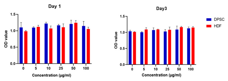
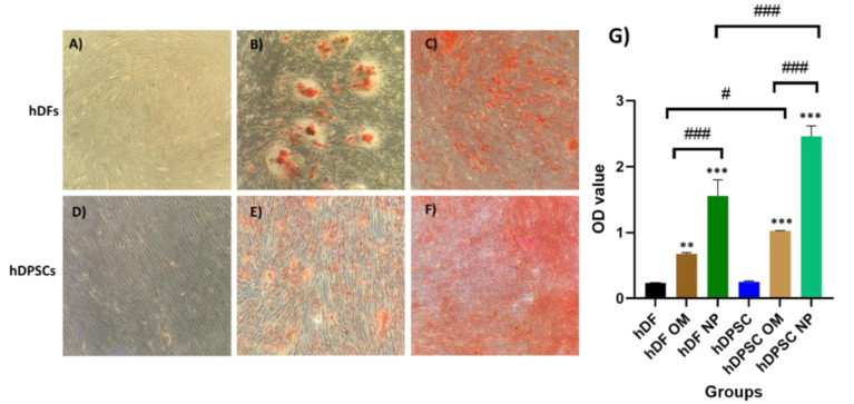
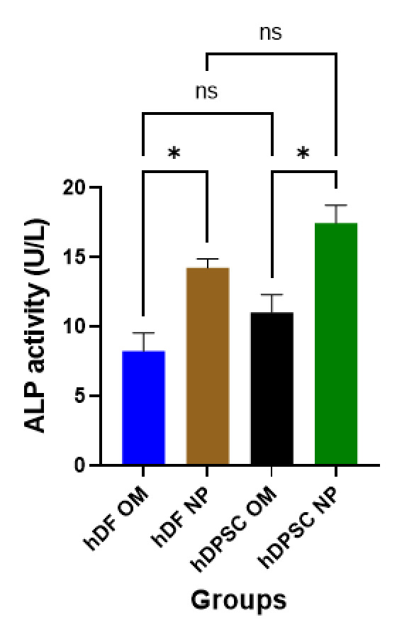
This study aimed to evaluate the osteogenic response of human dermal fibroblasts (hDFs) and human dental pulp stem cells (hDPSCs) when exposed to hydroxyapatite nanoparticles (HA-NPs), which are commonly employed biomaterials in the field of bone regeneration. The sol-gel method was employed to synthesize HA-NPs. Cell viability was assessed using the MTT assay after 24 and 72 hours of exposure to HA-NPs. Osteogenic differentiation was assessed through Alizarin red S staining (ARS), alkaline phosphatase (ALP) activity assay, and quantitative real-time PCR (qPCR) to evaluate the expression levels of osteogenic markers, including BMP-2, VEGF-A, RUNX2, and IL-6. The HA-NPs had a nanorod shape, with dimensions of 17-29 nm in width and 62-89 nm in length. Both hDPSCs and hDFs demonstrated enhanced osteogenic potential when exposed to HA-NPs, as indicated by increased ARS staining for calcium deposition and elevated ALP activity. Gene expression analysis showed up-regulation of BMP-2 and VEGF-A and down-regulation of RUNX2 in both cell types. IL-6 expression markedly increased in hDFs but did not show significant changes in hDPSCs compared to the control group. The findings of this study suggest that HA-NPs may enhance the osteogenic capability of hDPSCs and hDFs. The results demonstrate that while fibroblasts exhibit some mineralization potential, hDPSCs are the most suitable cell type for bone regenerative applications.
期刊介绍:
The International Journal of Molecular and Cellular Medicine (IJMCM) is a peer-reviewed, quarterly publication of Cellular and Molecular Biology Research Center (CMBRC), Babol University of Medical Sciences, Babol, Iran. The journal covers all cellular & molecular biology and medicine disciplines such as the genetic basis of disease, biomarker discovery in diagnosis and treatment, genomics and proteomics, bioinformatics, computer applications in human biology, stem cells and tissue engineering, medical biotechnology, nanomedicine, cellular processes related to growth, death and survival, clinical biochemistry, molecular & cellular immunology, molecular and cellular aspects of infectious disease and cancer research. IJMCM is a free access journal. All open access articles published in IJMCM are distributed under the terms of the Creative Commons Attribution CC BY. The journal doesn''t have any submission and article processing charges (APCs).

 求助内容:
求助内容: 应助结果提醒方式:


