Maximilian Feth, Mirabel Gracco, Michael Gröger, Melanie Hogg, Sandra Kress, Andrea Hoffmann, Enrico Calzia, Ulrich Wachter, Peter Radermacher, Tamara Merz
{"title":"硫代硫酸钠不影响小鼠外伤出血复苏期间的能量代谢或器官(日)功能。","authors":"Maximilian Feth, Mirabel Gracco, Michael Gröger, Melanie Hogg, Sandra Kress, Andrea Hoffmann, Enrico Calzia, Ulrich Wachter, Peter Radermacher, Tamara Merz","doi":"10.1186/s40635-025-00778-0","DOIUrl":null,"url":null,"abstract":"<p><strong>Background: </strong>In murine models, controversial data have been reported on the effect of hydrogen sulfide (H<sub>2</sub>S) administration during resuscitation from trauma-and-hemorrhage. The H<sub>2</sub>S donor sodium thiosulfate (Na<sub>2</sub>S<sub>2</sub>O<sub>3</sub>) is a recognized drug devoid of major side effects, and, hence, we determined its effects in our full scale ICU-model of resuscitated murine trauma-and-hemorrhage. We hypothesized that Na<sub>2</sub>S<sub>2</sub>O<sub>3</sub> might improve energy metabolism and thereby exert organ-protective effects as previously demonstrated in animals with genetic cystathionine-γ-lyase (CSE) deletion (CSE<sup>-/-</sup>).</p><p><strong>Methods: </strong>30 mice underwent combined blast wave-induced blunt chest trauma followed by 1 h of hemorrhagic shock (mean arterial pressure MAP = 35 ± 5 mmHg). Thereafter, resuscitation was initiated comprising re-transfusion of shed blood, lung-protective mechanical ventilation, fluid resuscitation and continuous i.v. noradrenaline infusion to maintain MAP > 55 mmHg over 6 h, and randomized administration of either i.v. 0.45 mg/g<sub>bodyweight</sub> Na<sub>2</sub>S<sub>2</sub>O<sub>3</sub> or vehicle (NaCl 0.9%). Hemodynamics, lung mechanics, gas exchange, acid-base-status and organ function parameters were recorded. Metabolic pathways were quantified based on gas chromatography/mass spectrometry assessment of plasma isotope enrichment during primed-continuous infusion of stable, non-radioactive, isotope labeled substrates. Mitochondrial function was determined using high-resolution respirometry, and tissue target proteins (nitrotyrosine formation, extravascular albumin accumulation, CSE expression) were analyzed using immunohistochemistry.</p><p><strong>Results: </strong>Data originate from 23 mice (Na<sub>2</sub>S<sub>2</sub>O<sub>3</sub> n = 12; vehicle n = 11)<sub>.</sub> Na<sub>2</sub>S<sub>2</sub>O<sub>3</sub> affected neither survival nor noradrenaline requirements. While minute ventilation had to be increased over time in both groups to maintain arterial PCO<sub>2</sub> without intergroup difference, arterial PO<sub>2</sub> decreased over time in Na<sub>2</sub>S<sub>2</sub>O<sub>3</sub>-treated mice (p = 0.006). Although arterial pH decreased in both groups (vehicle p = 0.049; Na<sub>2</sub>S<sub>2</sub>O<sub>3</sub> p < 0.001), metabolic acidosis was more pronounced in the Na<sub>2</sub>S<sub>2</sub>O<sub>3</sub> group. Neither metabolic pathways nor tissue mitochondrial respiratory activity or tissue target proteins showed any intergroup differences.</p><p><strong>Discussion: </strong>In this model of resuscitated trauma-and-hemorrhage, Na<sub>2</sub>S<sub>2</sub>O<sub>3</sub> did not exert any beneficial metabolic or organ-protective effect and was even associated with impaired pulmonary function. These results are in contrast to our previous findings in CSE<sup>-/-</sup> mice, but in line with more recent findings in CSE<sup>-/-</sup> mice with pre-existing comorbidities. Hence, our studies do not support a beneficial role of Na<sub>2</sub>S<sub>2</sub>O<sub>3</sub> in trauma resuscitation.</p>","PeriodicalId":13750,"journal":{"name":"Intensive Care Medicine Experimental","volume":"13 1","pages":"80"},"PeriodicalIF":2.8000,"publicationDate":"2025-08-06","publicationTypes":"Journal Article","fieldsOfStudy":null,"isOpenAccess":false,"openAccessPdf":"https://www.ncbi.nlm.nih.gov/pmc/articles/PMC12328856/pdf/","citationCount":"0","resultStr":"{\"title\":\"Sodium thiosulfate does not affect energy metabolism or organ (dys)function during resuscitation from murine trauma-and-hemorrhage.\",\"authors\":\"Maximilian Feth, Mirabel Gracco, Michael Gröger, Melanie Hogg, Sandra Kress, Andrea Hoffmann, Enrico Calzia, Ulrich Wachter, Peter Radermacher, Tamara Merz\",\"doi\":\"10.1186/s40635-025-00778-0\",\"DOIUrl\":null,\"url\":null,\"abstract\":\"<p><strong>Background: </strong>In murine models, controversial data have been reported on the effect of hydrogen sulfide (H<sub>2</sub>S) administration during resuscitation from trauma-and-hemorrhage. The H<sub>2</sub>S donor sodium thiosulfate (Na<sub>2</sub>S<sub>2</sub>O<sub>3</sub>) is a recognized drug devoid of major side effects, and, hence, we determined its effects in our full scale ICU-model of resuscitated murine trauma-and-hemorrhage. We hypothesized that Na<sub>2</sub>S<sub>2</sub>O<sub>3</sub> might improve energy metabolism and thereby exert organ-protective effects as previously demonstrated in animals with genetic cystathionine-γ-lyase (CSE) deletion (CSE<sup>-/-</sup>).</p><p><strong>Methods: </strong>30 mice underwent combined blast wave-induced blunt chest trauma followed by 1 h of hemorrhagic shock (mean arterial pressure MAP = 35 ± 5 mmHg). Thereafter, resuscitation was initiated comprising re-transfusion of shed blood, lung-protective mechanical ventilation, fluid resuscitation and continuous i.v. noradrenaline infusion to maintain MAP > 55 mmHg over 6 h, and randomized administration of either i.v. 0.45 mg/g<sub>bodyweight</sub> Na<sub>2</sub>S<sub>2</sub>O<sub>3</sub> or vehicle (NaCl 0.9%). Hemodynamics, lung mechanics, gas exchange, acid-base-status and organ function parameters were recorded. Metabolic pathways were quantified based on gas chromatography/mass spectrometry assessment of plasma isotope enrichment during primed-continuous infusion of stable, non-radioactive, isotope labeled substrates. Mitochondrial function was determined using high-resolution respirometry, and tissue target proteins (nitrotyrosine formation, extravascular albumin accumulation, CSE expression) were analyzed using immunohistochemistry.</p><p><strong>Results: </strong>Data originate from 23 mice (Na<sub>2</sub>S<sub>2</sub>O<sub>3</sub> n = 12; vehicle n = 11)<sub>.</sub> Na<sub>2</sub>S<sub>2</sub>O<sub>3</sub> affected neither survival nor noradrenaline requirements. While minute ventilation had to be increased over time in both groups to maintain arterial PCO<sub>2</sub> without intergroup difference, arterial PO<sub>2</sub> decreased over time in Na<sub>2</sub>S<sub>2</sub>O<sub>3</sub>-treated mice (p = 0.006). Although arterial pH decreased in both groups (vehicle p = 0.049; Na<sub>2</sub>S<sub>2</sub>O<sub>3</sub> p < 0.001), metabolic acidosis was more pronounced in the Na<sub>2</sub>S<sub>2</sub>O<sub>3</sub> group. Neither metabolic pathways nor tissue mitochondrial respiratory activity or tissue target proteins showed any intergroup differences.</p><p><strong>Discussion: </strong>In this model of resuscitated trauma-and-hemorrhage, Na<sub>2</sub>S<sub>2</sub>O<sub>3</sub> did not exert any beneficial metabolic or organ-protective effect and was even associated with impaired pulmonary function. These results are in contrast to our previous findings in CSE<sup>-/-</sup> mice, but in line with more recent findings in CSE<sup>-/-</sup> mice with pre-existing comorbidities. Hence, our studies do not support a beneficial role of Na<sub>2</sub>S<sub>2</sub>O<sub>3</sub> in trauma resuscitation.</p>\",\"PeriodicalId\":13750,\"journal\":{\"name\":\"Intensive Care Medicine Experimental\",\"volume\":\"13 1\",\"pages\":\"80\"},\"PeriodicalIF\":2.8000,\"publicationDate\":\"2025-08-06\",\"publicationTypes\":\"Journal Article\",\"fieldsOfStudy\":null,\"isOpenAccess\":false,\"openAccessPdf\":\"https://www.ncbi.nlm.nih.gov/pmc/articles/PMC12328856/pdf/\",\"citationCount\":\"0\",\"resultStr\":null,\"platform\":\"Semanticscholar\",\"paperid\":null,\"PeriodicalName\":\"Intensive Care Medicine Experimental\",\"FirstCategoryId\":\"1085\",\"ListUrlMain\":\"https://doi.org/10.1186/s40635-025-00778-0\",\"RegionNum\":0,\"RegionCategory\":null,\"ArticlePicture\":[],\"TitleCN\":null,\"AbstractTextCN\":null,\"PMCID\":null,\"EPubDate\":\"\",\"PubModel\":\"\",\"JCR\":\"Q2\",\"JCRName\":\"CRITICAL CARE MEDICINE\",\"Score\":null,\"Total\":0}","platform":"Semanticscholar","paperid":null,"PeriodicalName":"Intensive Care Medicine Experimental","FirstCategoryId":"1085","ListUrlMain":"https://doi.org/10.1186/s40635-025-00778-0","RegionNum":0,"RegionCategory":null,"ArticlePicture":[],"TitleCN":null,"AbstractTextCN":null,"PMCID":null,"EPubDate":"","PubModel":"","JCR":"Q2","JCRName":"CRITICAL CARE MEDICINE","Score":null,"Total":0}
Sodium thiosulfate does not affect energy metabolism or organ (dys)function during resuscitation from murine trauma-and-hemorrhage.
Background: In murine models, controversial data have been reported on the effect of hydrogen sulfide (H2S) administration during resuscitation from trauma-and-hemorrhage. The H2S donor sodium thiosulfate (Na2S2O3) is a recognized drug devoid of major side effects, and, hence, we determined its effects in our full scale ICU-model of resuscitated murine trauma-and-hemorrhage. We hypothesized that Na2S2O3 might improve energy metabolism and thereby exert organ-protective effects as previously demonstrated in animals with genetic cystathionine-γ-lyase (CSE) deletion (CSE-/-).
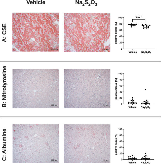
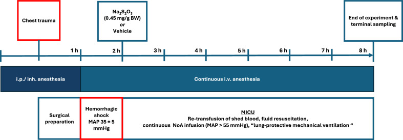
Methods: 30 mice underwent combined blast wave-induced blunt chest trauma followed by 1 h of hemorrhagic shock (mean arterial pressure MAP = 35 ± 5 mmHg). Thereafter, resuscitation was initiated comprising re-transfusion of shed blood, lung-protective mechanical ventilation, fluid resuscitation and continuous i.v. noradrenaline infusion to maintain MAP > 55 mmHg over 6 h, and randomized administration of either i.v. 0.45 mg/gbodyweight Na2S2O3 or vehicle (NaCl 0.9%). Hemodynamics, lung mechanics, gas exchange, acid-base-status and organ function parameters were recorded. Metabolic pathways were quantified based on gas chromatography/mass spectrometry assessment of plasma isotope enrichment during primed-continuous infusion of stable, non-radioactive, isotope labeled substrates. Mitochondrial function was determined using high-resolution respirometry, and tissue target proteins (nitrotyrosine formation, extravascular albumin accumulation, CSE expression) were analyzed using immunohistochemistry.
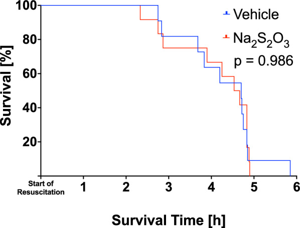
Results: Data originate from 23 mice (Na2S2O3 n = 12; vehicle n = 11). Na2S2O3 affected neither survival nor noradrenaline requirements. While minute ventilation had to be increased over time in both groups to maintain arterial PCO2 without intergroup difference, arterial PO2 decreased over time in Na2S2O3-treated mice (p = 0.006). Although arterial pH decreased in both groups (vehicle p = 0.049; Na2S2O3 p < 0.001), metabolic acidosis was more pronounced in the Na2S2O3 group. Neither metabolic pathways nor tissue mitochondrial respiratory activity or tissue target proteins showed any intergroup differences.
Discussion: In this model of resuscitated trauma-and-hemorrhage, Na2S2O3 did not exert any beneficial metabolic or organ-protective effect and was even associated with impaired pulmonary function. These results are in contrast to our previous findings in CSE-/- mice, but in line with more recent findings in CSE-/- mice with pre-existing comorbidities. Hence, our studies do not support a beneficial role of Na2S2O3 in trauma resuscitation.


 求助内容:
求助内容: 应助结果提醒方式:


