Rosemary Yeboah Ampofo, Kofi Adesi Kyei, Joseph Daniels, Andrew Yaw Nyantakyi
{"title":"在资源有限的情况下,确定沿z轴的冗余扫描覆盖范围和普通儿科计算机断层扫描成像检查的剂量含义。","authors":"Rosemary Yeboah Ampofo, Kofi Adesi Kyei, Joseph Daniels, Andrew Yaw Nyantakyi","doi":"10.1186/s12880-025-01860-5","DOIUrl":null,"url":null,"abstract":"<p><strong>Introduction: </strong>Optimizing scan parameters, particularly along the Z-axis, is crucial for minimizing unnecessary medical radiation exposure. Understanding and mitigating redundant scan coverages is essential for enhancing the safety and efficacy of paediatric CT imaging. This study quantified redundant Z-axis scan coverage and evaluated its associated dose implications in paediatric CT examinations in a limited-resource healthcare setting.</p><p><strong>Methods: </strong>A retrospective review was conducted on 279 paediatric CT scan records from two large tertiary hospitals in Ghana. Distances above the upper target and below the lower target of selected anatomical regions were measured using calibrated callipers on the CT console. The National Cancer Institute Dosimetry System for Computed Tomography (Monte Carlo-based software) was used to simulate the scanning situations and organ-dose implications.</p><p><strong>Results: </strong>In all, 87.3% of the CT scans had redundancies ranging from 0.77 ± 0.42 to 2.34 ± 1.22 cm for head, 1.43 ± 2.36 to 5.64 ± 3.00 cm for chest and 4.86 ± 3.01 to 6.75 ± 4.28 cm for abdominopelvic CT scans. Optimizing the scan length to the appropriate anatomical boundaries reduced the total dose-length product of neonates and middle-aged children by 27.9% and 26.1% respectively for chest CT scans, and 26.2% for infants during abdominopelvic CT scans. Optimal scan length selection for chest CT examinations resulted in organ-dose reductions of 57.5%, 56.1%, 63.6% and 73.8% for the thyroid glands, heart wall, lungs and breasts respectively.</p><p><strong>Conclusion: </strong>The study demonstrates a high prevalence (87.3%) of redundant scan coverage in paediatric CT examinations, with significant variances across different body sites. Implementing optimized scan lengths could substantially reduce radiation exposure and significantly lower organ doses.</p>","PeriodicalId":9020,"journal":{"name":"BMC Medical Imaging","volume":"25 1","pages":"317"},"PeriodicalIF":3.2000,"publicationDate":"2025-08-05","publicationTypes":"Journal Article","fieldsOfStudy":null,"isOpenAccess":false,"openAccessPdf":"https://www.ncbi.nlm.nih.gov/pmc/articles/PMC12326660/pdf/","citationCount":"0","resultStr":"{\"title\":\"Determination of redundant scan coverages along the Z-axis and dose implications for common paediatric computed tomography imaging examinations in a limited resource setting.\",\"authors\":\"Rosemary Yeboah Ampofo, Kofi Adesi Kyei, Joseph Daniels, Andrew Yaw Nyantakyi\",\"doi\":\"10.1186/s12880-025-01860-5\",\"DOIUrl\":null,\"url\":null,\"abstract\":\"<p><strong>Introduction: </strong>Optimizing scan parameters, particularly along the Z-axis, is crucial for minimizing unnecessary medical radiation exposure. Understanding and mitigating redundant scan coverages is essential for enhancing the safety and efficacy of paediatric CT imaging. This study quantified redundant Z-axis scan coverage and evaluated its associated dose implications in paediatric CT examinations in a limited-resource healthcare setting.</p><p><strong>Methods: </strong>A retrospective review was conducted on 279 paediatric CT scan records from two large tertiary hospitals in Ghana. Distances above the upper target and below the lower target of selected anatomical regions were measured using calibrated callipers on the CT console. The National Cancer Institute Dosimetry System for Computed Tomography (Monte Carlo-based software) was used to simulate the scanning situations and organ-dose implications.</p><p><strong>Results: </strong>In all, 87.3% of the CT scans had redundancies ranging from 0.77 ± 0.42 to 2.34 ± 1.22 cm for head, 1.43 ± 2.36 to 5.64 ± 3.00 cm for chest and 4.86 ± 3.01 to 6.75 ± 4.28 cm for abdominopelvic CT scans. Optimizing the scan length to the appropriate anatomical boundaries reduced the total dose-length product of neonates and middle-aged children by 27.9% and 26.1% respectively for chest CT scans, and 26.2% for infants during abdominopelvic CT scans. Optimal scan length selection for chest CT examinations resulted in organ-dose reductions of 57.5%, 56.1%, 63.6% and 73.8% for the thyroid glands, heart wall, lungs and breasts respectively.</p><p><strong>Conclusion: </strong>The study demonstrates a high prevalence (87.3%) of redundant scan coverage in paediatric CT examinations, with significant variances across different body sites. Implementing optimized scan lengths could substantially reduce radiation exposure and significantly lower organ doses.</p>\",\"PeriodicalId\":9020,\"journal\":{\"name\":\"BMC Medical Imaging\",\"volume\":\"25 1\",\"pages\":\"317\"},\"PeriodicalIF\":3.2000,\"publicationDate\":\"2025-08-05\",\"publicationTypes\":\"Journal Article\",\"fieldsOfStudy\":null,\"isOpenAccess\":false,\"openAccessPdf\":\"https://www.ncbi.nlm.nih.gov/pmc/articles/PMC12326660/pdf/\",\"citationCount\":\"0\",\"resultStr\":null,\"platform\":\"Semanticscholar\",\"paperid\":null,\"PeriodicalName\":\"BMC Medical Imaging\",\"FirstCategoryId\":\"3\",\"ListUrlMain\":\"https://doi.org/10.1186/s12880-025-01860-5\",\"RegionNum\":3,\"RegionCategory\":\"医学\",\"ArticlePicture\":[],\"TitleCN\":null,\"AbstractTextCN\":null,\"PMCID\":null,\"EPubDate\":\"\",\"PubModel\":\"\",\"JCR\":\"Q2\",\"JCRName\":\"RADIOLOGY, NUCLEAR MEDICINE & MEDICAL IMAGING\",\"Score\":null,\"Total\":0}","platform":"Semanticscholar","paperid":null,"PeriodicalName":"BMC Medical Imaging","FirstCategoryId":"3","ListUrlMain":"https://doi.org/10.1186/s12880-025-01860-5","RegionNum":3,"RegionCategory":"医学","ArticlePicture":[],"TitleCN":null,"AbstractTextCN":null,"PMCID":null,"EPubDate":"","PubModel":"","JCR":"Q2","JCRName":"RADIOLOGY, NUCLEAR MEDICINE & MEDICAL IMAGING","Score":null,"Total":0}
Determination of redundant scan coverages along the Z-axis and dose implications for common paediatric computed tomography imaging examinations in a limited resource setting.
Introduction: Optimizing scan parameters, particularly along the Z-axis, is crucial for minimizing unnecessary medical radiation exposure. Understanding and mitigating redundant scan coverages is essential for enhancing the safety and efficacy of paediatric CT imaging. This study quantified redundant Z-axis scan coverage and evaluated its associated dose implications in paediatric CT examinations in a limited-resource healthcare setting.
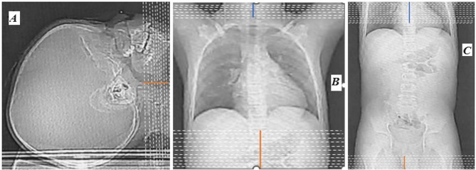
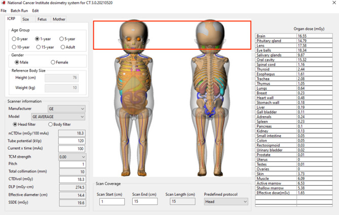
Methods: A retrospective review was conducted on 279 paediatric CT scan records from two large tertiary hospitals in Ghana. Distances above the upper target and below the lower target of selected anatomical regions were measured using calibrated callipers on the CT console. The National Cancer Institute Dosimetry System for Computed Tomography (Monte Carlo-based software) was used to simulate the scanning situations and organ-dose implications.
Results: In all, 87.3% of the CT scans had redundancies ranging from 0.77 ± 0.42 to 2.34 ± 1.22 cm for head, 1.43 ± 2.36 to 5.64 ± 3.00 cm for chest and 4.86 ± 3.01 to 6.75 ± 4.28 cm for abdominopelvic CT scans. Optimizing the scan length to the appropriate anatomical boundaries reduced the total dose-length product of neonates and middle-aged children by 27.9% and 26.1% respectively for chest CT scans, and 26.2% for infants during abdominopelvic CT scans. Optimal scan length selection for chest CT examinations resulted in organ-dose reductions of 57.5%, 56.1%, 63.6% and 73.8% for the thyroid glands, heart wall, lungs and breasts respectively.
Conclusion: The study demonstrates a high prevalence (87.3%) of redundant scan coverage in paediatric CT examinations, with significant variances across different body sites. Implementing optimized scan lengths could substantially reduce radiation exposure and significantly lower organ doses.
期刊介绍:
BMC Medical Imaging is an open access journal publishing original peer-reviewed research articles in the development, evaluation, and use of imaging techniques and image processing tools to diagnose and manage disease.


 求助内容:
求助内容: 应助结果提醒方式:


