M Madhuvandhi, K Sankar, Latha Asokan, S Duraimurugan
{"title":"后上颌骨中央巨细胞肉芽肿1例。","authors":"M Madhuvandhi, K Sankar, Latha Asokan, S Duraimurugan","doi":"10.4103/ams.ams_130_24","DOIUrl":null,"url":null,"abstract":"<p><strong>Rationale: </strong>Central giant cell granuloma (CGCG) is a non-neoplastic proliferative lesion that clinically and radiologically mimics various lesions without any unique pathognomic features.</p><p><strong>Patient concerns: </strong>A 14-year-old male patient reported with a chief complaint of swelling in the right upper back tooth region for four months.</p><p><strong>Diagnosis: </strong>Incisional biopsy of the lesion shows numerous dilated and thin-walled blood vessels and focal areas of chronic inflammatory cell infiltrate beneath the epithelium suggestive of angiofibroma.</p><p><strong>Treatment: </strong>Surgical excision of the lesion along with curettage was done under general anaesthesia. On histopathologic examination, the lesion revealed loose connective tissue stroma with mesenchymal cells and multinucleated giant cells suggestive of CGCG.</p><p><strong>Outcomes: </strong>The patient was followed up for one year without any evidence of recurrences.</p><p><strong>Take-away lessons: </strong>Presentation of CGCG of the oral cavity is rare and mimics a wide variety of lesions clinically and radiologically. Histopathology remains the only diagnostic modality.</p>","PeriodicalId":7972,"journal":{"name":"Annals of Maxillofacial Surgery","volume":"15 1","pages":"95-97"},"PeriodicalIF":0.0000,"publicationDate":"2025-01-01","publicationTypes":"Journal Article","fieldsOfStudy":null,"isOpenAccess":false,"openAccessPdf":"https://www.ncbi.nlm.nih.gov/pmc/articles/PMC12321165/pdf/","citationCount":"0","resultStr":"{\"title\":\"Central Giant Cell Granuloma of Posterior Maxilla - A Case Report.\",\"authors\":\"M Madhuvandhi, K Sankar, Latha Asokan, S Duraimurugan\",\"doi\":\"10.4103/ams.ams_130_24\",\"DOIUrl\":null,\"url\":null,\"abstract\":\"<p><strong>Rationale: </strong>Central giant cell granuloma (CGCG) is a non-neoplastic proliferative lesion that clinically and radiologically mimics various lesions without any unique pathognomic features.</p><p><strong>Patient concerns: </strong>A 14-year-old male patient reported with a chief complaint of swelling in the right upper back tooth region for four months.</p><p><strong>Diagnosis: </strong>Incisional biopsy of the lesion shows numerous dilated and thin-walled blood vessels and focal areas of chronic inflammatory cell infiltrate beneath the epithelium suggestive of angiofibroma.</p><p><strong>Treatment: </strong>Surgical excision of the lesion along with curettage was done under general anaesthesia. On histopathologic examination, the lesion revealed loose connective tissue stroma with mesenchymal cells and multinucleated giant cells suggestive of CGCG.</p><p><strong>Outcomes: </strong>The patient was followed up for one year without any evidence of recurrences.</p><p><strong>Take-away lessons: </strong>Presentation of CGCG of the oral cavity is rare and mimics a wide variety of lesions clinically and radiologically. Histopathology remains the only diagnostic modality.</p>\",\"PeriodicalId\":7972,\"journal\":{\"name\":\"Annals of Maxillofacial Surgery\",\"volume\":\"15 1\",\"pages\":\"95-97\"},\"PeriodicalIF\":0.0000,\"publicationDate\":\"2025-01-01\",\"publicationTypes\":\"Journal Article\",\"fieldsOfStudy\":null,\"isOpenAccess\":false,\"openAccessPdf\":\"https://www.ncbi.nlm.nih.gov/pmc/articles/PMC12321165/pdf/\",\"citationCount\":\"0\",\"resultStr\":null,\"platform\":\"Semanticscholar\",\"paperid\":null,\"PeriodicalName\":\"Annals of Maxillofacial Surgery\",\"FirstCategoryId\":\"1085\",\"ListUrlMain\":\"https://doi.org/10.4103/ams.ams_130_24\",\"RegionNum\":0,\"RegionCategory\":null,\"ArticlePicture\":[],\"TitleCN\":null,\"AbstractTextCN\":null,\"PMCID\":null,\"EPubDate\":\"2025/3/18 0:00:00\",\"PubModel\":\"Epub\",\"JCR\":\"Q2\",\"JCRName\":\"Dentistry\",\"Score\":null,\"Total\":0}","platform":"Semanticscholar","paperid":null,"PeriodicalName":"Annals of Maxillofacial Surgery","FirstCategoryId":"1085","ListUrlMain":"https://doi.org/10.4103/ams.ams_130_24","RegionNum":0,"RegionCategory":null,"ArticlePicture":[],"TitleCN":null,"AbstractTextCN":null,"PMCID":null,"EPubDate":"2025/3/18 0:00:00","PubModel":"Epub","JCR":"Q2","JCRName":"Dentistry","Score":null,"Total":0}
Central Giant Cell Granuloma of Posterior Maxilla - A Case Report.
Rationale: Central giant cell granuloma (CGCG) is a non-neoplastic proliferative lesion that clinically and radiologically mimics various lesions without any unique pathognomic features.
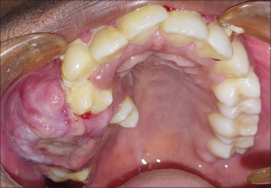
Patient concerns: A 14-year-old male patient reported with a chief complaint of swelling in the right upper back tooth region for four months.
Diagnosis: Incisional biopsy of the lesion shows numerous dilated and thin-walled blood vessels and focal areas of chronic inflammatory cell infiltrate beneath the epithelium suggestive of angiofibroma.
Treatment: Surgical excision of the lesion along with curettage was done under general anaesthesia. On histopathologic examination, the lesion revealed loose connective tissue stroma with mesenchymal cells and multinucleated giant cells suggestive of CGCG.
Outcomes: The patient was followed up for one year without any evidence of recurrences.
Take-away lessons: Presentation of CGCG of the oral cavity is rare and mimics a wide variety of lesions clinically and radiologically. Histopathology remains the only diagnostic modality.

 求助内容:
求助内容: 应助结果提醒方式:


