{"title":"增强现实在颞下颌关节置换术中的应用-一项评估性临床研究。","authors":"Jigna S Shah, Manish K Poojary","doi":"10.4103/ams.ams_45_24","DOIUrl":null,"url":null,"abstract":"<p><strong>Introduction: </strong>Augmented reality (AR) combines computer-generated images, audios and videos on a screen with real-life scenes. Arthrocentesis is the lavage of the temporomandibular joint (TMJ) using sterile needles and sterile irrigants which requires precise puncture by a needle to the upper joint compartment. The aim of the study was to develop an AR system to identify the puncture point for TMJ arthrocentesis with direct access to the upper compartment of the joint.</p><p><strong>Materials and methods: </strong>Two cases diagnosed with chronic recurrent dislocation of TMJ were selected for the study. A three-dimensional virtual model was reconstructed from cone-beam computed tomography (CBCT) images of a patient. The AR visualisation was created by developing an Android app which superimposed the virtual model and the TMJ in real time. The skin puncture point was marked on the skin, and the needle was guided to the upper compartment of the joint using this superimposed view to perform TMJ arthrocentesis.</p><p><strong>Results: </strong>Puncture with the AR method was successful in both the cases with direct access to the upper compartment of TMJ. Pain had reduced completely in both cases during 30- and 60-day follow-up periods; maximum mouth opening had reduced by an average of 4.33 mm after 60 days postoperatively, and clicking sound was completely absent after 14-day follow-up period.</p><p><strong>Discussion: </strong>AR could potentially enhance the accuracy and effectiveness of TMJ arthrocentesis by providing precise needle guidance and reducing the need for multiple punctures. However, the limitations must be addressed as well.</p>","PeriodicalId":7972,"journal":{"name":"Annals of Maxillofacial Surgery","volume":"15 1","pages":"78-82"},"PeriodicalIF":0.0000,"publicationDate":"2025-01-01","publicationTypes":"Journal Article","fieldsOfStudy":null,"isOpenAccess":false,"openAccessPdf":"https://www.ncbi.nlm.nih.gov/pmc/articles/PMC12321183/pdf/","citationCount":"0","resultStr":"{\"title\":\"Augmented Reality in Temporomandibular Joint Arthrocentesis - An Evaluative Clinical Study.\",\"authors\":\"Jigna S Shah, Manish K Poojary\",\"doi\":\"10.4103/ams.ams_45_24\",\"DOIUrl\":null,\"url\":null,\"abstract\":\"<p><strong>Introduction: </strong>Augmented reality (AR) combines computer-generated images, audios and videos on a screen with real-life scenes. Arthrocentesis is the lavage of the temporomandibular joint (TMJ) using sterile needles and sterile irrigants which requires precise puncture by a needle to the upper joint compartment. The aim of the study was to develop an AR system to identify the puncture point for TMJ arthrocentesis with direct access to the upper compartment of the joint.</p><p><strong>Materials and methods: </strong>Two cases diagnosed with chronic recurrent dislocation of TMJ were selected for the study. A three-dimensional virtual model was reconstructed from cone-beam computed tomography (CBCT) images of a patient. The AR visualisation was created by developing an Android app which superimposed the virtual model and the TMJ in real time. The skin puncture point was marked on the skin, and the needle was guided to the upper compartment of the joint using this superimposed view to perform TMJ arthrocentesis.</p><p><strong>Results: </strong>Puncture with the AR method was successful in both the cases with direct access to the upper compartment of TMJ. Pain had reduced completely in both cases during 30- and 60-day follow-up periods; maximum mouth opening had reduced by an average of 4.33 mm after 60 days postoperatively, and clicking sound was completely absent after 14-day follow-up period.</p><p><strong>Discussion: </strong>AR could potentially enhance the accuracy and effectiveness of TMJ arthrocentesis by providing precise needle guidance and reducing the need for multiple punctures. However, the limitations must be addressed as well.</p>\",\"PeriodicalId\":7972,\"journal\":{\"name\":\"Annals of Maxillofacial Surgery\",\"volume\":\"15 1\",\"pages\":\"78-82\"},\"PeriodicalIF\":0.0000,\"publicationDate\":\"2025-01-01\",\"publicationTypes\":\"Journal Article\",\"fieldsOfStudy\":null,\"isOpenAccess\":false,\"openAccessPdf\":\"https://www.ncbi.nlm.nih.gov/pmc/articles/PMC12321183/pdf/\",\"citationCount\":\"0\",\"resultStr\":null,\"platform\":\"Semanticscholar\",\"paperid\":null,\"PeriodicalName\":\"Annals of Maxillofacial Surgery\",\"FirstCategoryId\":\"1085\",\"ListUrlMain\":\"https://doi.org/10.4103/ams.ams_45_24\",\"RegionNum\":0,\"RegionCategory\":null,\"ArticlePicture\":[],\"TitleCN\":null,\"AbstractTextCN\":null,\"PMCID\":null,\"EPubDate\":\"2025/4/29 0:00:00\",\"PubModel\":\"Epub\",\"JCR\":\"Q2\",\"JCRName\":\"Dentistry\",\"Score\":null,\"Total\":0}","platform":"Semanticscholar","paperid":null,"PeriodicalName":"Annals of Maxillofacial Surgery","FirstCategoryId":"1085","ListUrlMain":"https://doi.org/10.4103/ams.ams_45_24","RegionNum":0,"RegionCategory":null,"ArticlePicture":[],"TitleCN":null,"AbstractTextCN":null,"PMCID":null,"EPubDate":"2025/4/29 0:00:00","PubModel":"Epub","JCR":"Q2","JCRName":"Dentistry","Score":null,"Total":0}
Augmented Reality in Temporomandibular Joint Arthrocentesis - An Evaluative Clinical Study.
Introduction: Augmented reality (AR) combines computer-generated images, audios and videos on a screen with real-life scenes. Arthrocentesis is the lavage of the temporomandibular joint (TMJ) using sterile needles and sterile irrigants which requires precise puncture by a needle to the upper joint compartment. The aim of the study was to develop an AR system to identify the puncture point for TMJ arthrocentesis with direct access to the upper compartment of the joint.
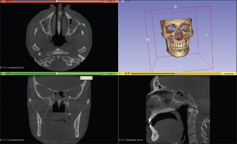
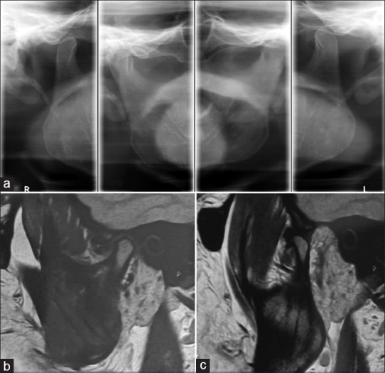
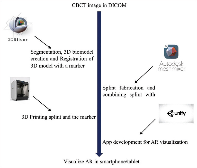
Materials and methods: Two cases diagnosed with chronic recurrent dislocation of TMJ were selected for the study. A three-dimensional virtual model was reconstructed from cone-beam computed tomography (CBCT) images of a patient. The AR visualisation was created by developing an Android app which superimposed the virtual model and the TMJ in real time. The skin puncture point was marked on the skin, and the needle was guided to the upper compartment of the joint using this superimposed view to perform TMJ arthrocentesis.
Results: Puncture with the AR method was successful in both the cases with direct access to the upper compartment of TMJ. Pain had reduced completely in both cases during 30- and 60-day follow-up periods; maximum mouth opening had reduced by an average of 4.33 mm after 60 days postoperatively, and clicking sound was completely absent after 14-day follow-up period.
Discussion: AR could potentially enhance the accuracy and effectiveness of TMJ arthrocentesis by providing precise needle guidance and reducing the need for multiple punctures. However, the limitations must be addressed as well.

 求助内容:
求助内容: 应助结果提醒方式:


