Ying Liu, Jiake Mo, Zi Guo, Jiaqi Zhang, Weian Tang, Xubiao Meng, Yufang Luo, Fang Wang, Zhaohui Mo
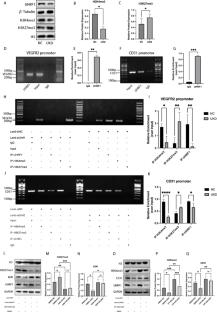
{"title":"内皮细胞中的UHRF1对血管生成至关重要,并与促血管生成信号通路的激活和内皮基因的表达有关。","authors":"Ying Liu, Jiake Mo, Zi Guo, Jiaqi Zhang, Weian Tang, Xubiao Meng, Yufang Luo, Fang Wang, Zhaohui Mo","doi":"10.1007/s10456-025-09998-0","DOIUrl":null,"url":null,"abstract":"<div><p>Epigenetics is increasingly recognized as a crucial factor in angiogenesis. Ubiquitin-like with PHD and RING Finger Domains 1 (UHRF1) is an important epigenetic regulatory protein involved in regulating cellular life processes, developing many diseases. However, its potential role in regulating embryonic vascular development and postnatal angiogenesis is unclear. Our study found that endothelial cell-specific UHRF1 knockout mice showed obvious developmental disorders at the embryonic stage (E11.5-15.5), including impaired development of the individual embryo size and organs, sparse vascularity in the yolk sac, or even death. In the lower limb ischemia model, UHRF1 expression in ischemic muscle tissues of mice is proportionate to the regeneration of blood vessels. To confirm the specific inhibition of UHRF1, we transfected an adeno-associated virus serotype 9 which inserted a TIE-2 promoter and mediated the delivery of short hairpin RNA (AAV9-TIE-2-shUHRF1) into mouse vascular endothelial cells to knock down UHRF1 specifically. We observed that the knockdown of UHRF1 in endothelial cells results in poorer lower limb perfusion in mice. Mechanically, UHRF1 knockdown decreased the tube-forming capacity of ECFCs, whereas overexpression of UHRF1 by diabetic ECFCs where UHRF1 expression is typically downregulated significantly increased the tube-forming capacity of the cells. RNAseq and related bioinformatics analyses showed that differentially expressed genes (DEGs) were mainly involved in angiogenesis-related pathways. The results of qPCR and western blot showed that the protein and mRNA levels of angiogenesis-related factors (VEGF, PDGF, and ANGPT1), as well as vascular endothelial surface marker molecules (VEGFR2, CD31, and c-Kit), were down-regulated accordingly. Furthermore, ChIP experiments showed that UHRF1 was able to bind the promoters of VEGFR2 and CD31, affecting the levels of histone-methylated protein (H3K4me3 and H3K27me3) enriched in the promoter region. However, the expression of CD31 and VEGFR2 can be reversed separately after the transformation of different histone-methylated protein levels (H3K4me3 and H3K27me3). Taken together, UHRF1 may regulate angiogenic gene expression and vascular endothelial cell differentiation through epigenetic mechanisms and is essential for angiogenesis.</p></div>","PeriodicalId":7886,"journal":{"name":"Angiogenesis","volume":"28 4","pages":""},"PeriodicalIF":9.2000,"publicationDate":"2025-08-05","publicationTypes":"Journal Article","fieldsOfStudy":null,"isOpenAccess":false,"openAccessPdf":"","citationCount":"0","resultStr":"{\"title\":\"UHRF1 in endothelial cells is essential for angiogenesis and associated with the activation of pro-angiogenic signaling pathways and expression of endothelial genes\",\"authors\":\"Ying Liu, Jiake Mo, Zi Guo, Jiaqi Zhang, Weian Tang, Xubiao Meng, Yufang Luo, Fang Wang, Zhaohui Mo\",\"doi\":\"10.1007/s10456-025-09998-0\",\"DOIUrl\":null,\"url\":null,\"abstract\":\"<div><p>Epigenetics is increasingly recognized as a crucial factor in angiogenesis. Ubiquitin-like with PHD and RING Finger Domains 1 (UHRF1) is an important epigenetic regulatory protein involved in regulating cellular life processes, developing many diseases. However, its potential role in regulating embryonic vascular development and postnatal angiogenesis is unclear. Our study found that endothelial cell-specific UHRF1 knockout mice showed obvious developmental disorders at the embryonic stage (E11.5-15.5), including impaired development of the individual embryo size and organs, sparse vascularity in the yolk sac, or even death. In the lower limb ischemia model, UHRF1 expression in ischemic muscle tissues of mice is proportionate to the regeneration of blood vessels. To confirm the specific inhibition of UHRF1, we transfected an adeno-associated virus serotype 9 which inserted a TIE-2 promoter and mediated the delivery of short hairpin RNA (AAV9-TIE-2-shUHRF1) into mouse vascular endothelial cells to knock down UHRF1 specifically. We observed that the knockdown of UHRF1 in endothelial cells results in poorer lower limb perfusion in mice. Mechanically, UHRF1 knockdown decreased the tube-forming capacity of ECFCs, whereas overexpression of UHRF1 by diabetic ECFCs where UHRF1 expression is typically downregulated significantly increased the tube-forming capacity of the cells. RNAseq and related bioinformatics analyses showed that differentially expressed genes (DEGs) were mainly involved in angiogenesis-related pathways. The results of qPCR and western blot showed that the protein and mRNA levels of angiogenesis-related factors (VEGF, PDGF, and ANGPT1), as well as vascular endothelial surface marker molecules (VEGFR2, CD31, and c-Kit), were down-regulated accordingly. Furthermore, ChIP experiments showed that UHRF1 was able to bind the promoters of VEGFR2 and CD31, affecting the levels of histone-methylated protein (H3K4me3 and H3K27me3) enriched in the promoter region. However, the expression of CD31 and VEGFR2 can be reversed separately after the transformation of different histone-methylated protein levels (H3K4me3 and H3K27me3). Taken together, UHRF1 may regulate angiogenic gene expression and vascular endothelial cell differentiation through epigenetic mechanisms and is essential for angiogenesis.</p></div>\",\"PeriodicalId\":7886,\"journal\":{\"name\":\"Angiogenesis\",\"volume\":\"28 4\",\"pages\":\"\"},\"PeriodicalIF\":9.2000,\"publicationDate\":\"2025-08-05\",\"publicationTypes\":\"Journal Article\",\"fieldsOfStudy\":null,\"isOpenAccess\":false,\"openAccessPdf\":\"\",\"citationCount\":\"0\",\"resultStr\":null,\"platform\":\"Semanticscholar\",\"paperid\":null,\"PeriodicalName\":\"Angiogenesis\",\"FirstCategoryId\":\"3\",\"ListUrlMain\":\"https://link.springer.com/article/10.1007/s10456-025-09998-0\",\"RegionNum\":1,\"RegionCategory\":\"医学\",\"ArticlePicture\":[],\"TitleCN\":null,\"AbstractTextCN\":null,\"PMCID\":null,\"EPubDate\":\"\",\"PubModel\":\"\",\"JCR\":\"Q1\",\"JCRName\":\"PERIPHERAL VASCULAR DISEASE\",\"Score\":null,\"Total\":0}","platform":"Semanticscholar","paperid":null,"PeriodicalName":"Angiogenesis","FirstCategoryId":"3","ListUrlMain":"https://link.springer.com/article/10.1007/s10456-025-09998-0","RegionNum":1,"RegionCategory":"医学","ArticlePicture":[],"TitleCN":null,"AbstractTextCN":null,"PMCID":null,"EPubDate":"","PubModel":"","JCR":"Q1","JCRName":"PERIPHERAL VASCULAR DISEASE","Score":null,"Total":0}
引用次数: 0
摘要
表观遗传学越来越被认为是血管生成的关键因素。泛素样蛋白(Ubiquitin-like with PHD and RING Finger Domains 1, UHRF1)是一种重要的表观遗传调控蛋白,参与细胞生命过程的调控,参与多种疾病的发生。然而,其在调节胚胎血管发育和出生后血管生成中的潜在作用尚不清楚。我们的研究发现内皮细胞特异性UHRF1基因敲除小鼠在胚胎期(E11.5-15.5)表现出明显的发育障碍,包括个体胚胎大小和器官发育受损,卵黄囊血管稀疏,甚至死亡。在下肢缺血模型中,UHRF1在小鼠缺血肌肉组织中的表达与血管再生成正比。为了证实UHRF1的特异性抑制作用,我们转染了一种血清型9型腺相关病毒,该病毒插入TIE-2启动子,介导短发夹RNA (AAV9-TIE-2-shUHRF1)传递到小鼠血管内皮细胞中,特异性地敲除UHRF1。我们观察到内皮细胞中UHRF1的敲低导致小鼠下肢灌注变差。从机械上讲,UHRF1敲低会降低ecfc的成管能力,而UHRF1表达通常下调的糖尿病ecfc过表达UHRF1会显著增加细胞的成管能力。RNAseq和相关生物信息学分析表明,差异表达基因(DEGs)主要参与血管生成相关途径。qPCR和western blot结果显示,血管生成相关因子(VEGF、PDGF、ANGPT1)和血管内皮表面标记分子(VEGFR2、CD31、c-Kit)的蛋白和mRNA水平相应下调。此外,ChIP实验表明,UHRF1能够结合VEGFR2和CD31的启动子,影响启动子区域富集的组蛋白甲基化蛋白(H3K4me3和H3K27me3)的水平。然而,不同组蛋白甲基化蛋白水平(H3K4me3和H3K27me3)转化后,CD31和VEGFR2的表达可以分别逆转。综上所述,UHRF1可能通过表观遗传机制调控血管生成基因表达和血管内皮细胞分化,对血管生成至关重要。
UHRF1 in endothelial cells is essential for angiogenesis and associated with the activation of pro-angiogenic signaling pathways and expression of endothelial genes
Epigenetics is increasingly recognized as a crucial factor in angiogenesis. Ubiquitin-like with PHD and RING Finger Domains 1 (UHRF1) is an important epigenetic regulatory protein involved in regulating cellular life processes, developing many diseases. However, its potential role in regulating embryonic vascular development and postnatal angiogenesis is unclear. Our study found that endothelial cell-specific UHRF1 knockout mice showed obvious developmental disorders at the embryonic stage (E11.5-15.5), including impaired development of the individual embryo size and organs, sparse vascularity in the yolk sac, or even death. In the lower limb ischemia model, UHRF1 expression in ischemic muscle tissues of mice is proportionate to the regeneration of blood vessels. To confirm the specific inhibition of UHRF1, we transfected an adeno-associated virus serotype 9 which inserted a TIE-2 promoter and mediated the delivery of short hairpin RNA (AAV9-TIE-2-shUHRF1) into mouse vascular endothelial cells to knock down UHRF1 specifically. We observed that the knockdown of UHRF1 in endothelial cells results in poorer lower limb perfusion in mice. Mechanically, UHRF1 knockdown decreased the tube-forming capacity of ECFCs, whereas overexpression of UHRF1 by diabetic ECFCs where UHRF1 expression is typically downregulated significantly increased the tube-forming capacity of the cells. RNAseq and related bioinformatics analyses showed that differentially expressed genes (DEGs) were mainly involved in angiogenesis-related pathways. The results of qPCR and western blot showed that the protein and mRNA levels of angiogenesis-related factors (VEGF, PDGF, and ANGPT1), as well as vascular endothelial surface marker molecules (VEGFR2, CD31, and c-Kit), were down-regulated accordingly. Furthermore, ChIP experiments showed that UHRF1 was able to bind the promoters of VEGFR2 and CD31, affecting the levels of histone-methylated protein (H3K4me3 and H3K27me3) enriched in the promoter region. However, the expression of CD31 and VEGFR2 can be reversed separately after the transformation of different histone-methylated protein levels (H3K4me3 and H3K27me3). Taken together, UHRF1 may regulate angiogenic gene expression and vascular endothelial cell differentiation through epigenetic mechanisms and is essential for angiogenesis.
期刊介绍:
Angiogenesis, a renowned international journal, seeks to publish high-quality original articles and reviews on the cellular and molecular mechanisms governing angiogenesis in both normal and pathological conditions. By serving as a primary platform for swift communication within the field of angiogenesis research, this multidisciplinary journal showcases pioneering experimental studies utilizing molecular techniques, in vitro methods, animal models, and clinical investigations into angiogenic diseases. Furthermore, Angiogenesis sheds light on cutting-edge therapeutic strategies for promoting or inhibiting angiogenesis, while also highlighting fresh markers and techniques for disease diagnosis and prognosis.
 求助内容:
求助内容: 应助结果提醒方式:


