{"title":"上颌副窦口的患病率及其性别和亚型分布的回顾性研究。","authors":"Shams Ul Nisa, Aishwarya Umesh Lohokare, Shameeka Thopte, Samir Dashrath Khaire, Neelam Gavali, Kinjal Shankar Lakhani","doi":"10.4103/ams.ams_148_24","DOIUrl":null,"url":null,"abstract":"<p><strong>Introduction: </strong>This epidemiological retrospective study evaluated cone beam computed tomography (CBCT) as a reliable and valid tool in assessing maxillary sinus (MS) morphology, location and prevalence of accessory maxillary ostia (AMO) and evaluating its distribution with regards to gender and its subtypes.</p><p><strong>Materials and methods: </strong>The reporting of the study was done in accordance to strengthening the reporting of observational studies in epidemiology guidelines/checklist. Two hundred CBCT scans (400 MSs) were analysed for height and width of the maxillary sinuses (MSs) and its distribution with regards to gender. The prevalence of AMO and its different types of shapes were evaluated.</p><p><strong>Results: </strong>Between the male and female groups, differences on both the right and left sides with respect to the MS height were observed (<i>P</i> > 0.05). However, the male group showed statistically significant higher values for right-sided MS width (<i>P</i> < 0.05). The prevalence of AMO in 400 MSs was found to be 65%, of which 63% of AMOs were present in the male group. The prevalence of types of accessory maxillary ostia revealed that the round and ovoid shapes were similar in number as compared to slit shapes.</p><p><strong>Discussion: </strong>The forensic anthropology through CBCT can utilise the MS width as a tool to assess various anatomical variations. The frequency of AMOs can be utilised as a critical indicator for assessing such morphological and anatomical variations. As a result, the excellent spatial resolution of CBCT enables it as a reliable tool for identification of even smallest anatomical variations in MS.</p>","PeriodicalId":7972,"journal":{"name":"Annals of Maxillofacial Surgery","volume":"15 1","pages":"67-71"},"PeriodicalIF":0.0000,"publicationDate":"2025-01-01","publicationTypes":"Journal Article","fieldsOfStudy":null,"isOpenAccess":false,"openAccessPdf":"https://www.ncbi.nlm.nih.gov/pmc/articles/PMC12321187/pdf/","citationCount":"0","resultStr":"{\"title\":\"Prevalence of Accessory Maxillary Sinus Ostia and its Distribution with Gender and Subtypes - A Retrospective Study.\",\"authors\":\"Shams Ul Nisa, Aishwarya Umesh Lohokare, Shameeka Thopte, Samir Dashrath Khaire, Neelam Gavali, Kinjal Shankar Lakhani\",\"doi\":\"10.4103/ams.ams_148_24\",\"DOIUrl\":null,\"url\":null,\"abstract\":\"<p><strong>Introduction: </strong>This epidemiological retrospective study evaluated cone beam computed tomography (CBCT) as a reliable and valid tool in assessing maxillary sinus (MS) morphology, location and prevalence of accessory maxillary ostia (AMO) and evaluating its distribution with regards to gender and its subtypes.</p><p><strong>Materials and methods: </strong>The reporting of the study was done in accordance to strengthening the reporting of observational studies in epidemiology guidelines/checklist. Two hundred CBCT scans (400 MSs) were analysed for height and width of the maxillary sinuses (MSs) and its distribution with regards to gender. The prevalence of AMO and its different types of shapes were evaluated.</p><p><strong>Results: </strong>Between the male and female groups, differences on both the right and left sides with respect to the MS height were observed (<i>P</i> > 0.05). However, the male group showed statistically significant higher values for right-sided MS width (<i>P</i> < 0.05). The prevalence of AMO in 400 MSs was found to be 65%, of which 63% of AMOs were present in the male group. The prevalence of types of accessory maxillary ostia revealed that the round and ovoid shapes were similar in number as compared to slit shapes.</p><p><strong>Discussion: </strong>The forensic anthropology through CBCT can utilise the MS width as a tool to assess various anatomical variations. The frequency of AMOs can be utilised as a critical indicator for assessing such morphological and anatomical variations. As a result, the excellent spatial resolution of CBCT enables it as a reliable tool for identification of even smallest anatomical variations in MS.</p>\",\"PeriodicalId\":7972,\"journal\":{\"name\":\"Annals of Maxillofacial Surgery\",\"volume\":\"15 1\",\"pages\":\"67-71\"},\"PeriodicalIF\":0.0000,\"publicationDate\":\"2025-01-01\",\"publicationTypes\":\"Journal Article\",\"fieldsOfStudy\":null,\"isOpenAccess\":false,\"openAccessPdf\":\"https://www.ncbi.nlm.nih.gov/pmc/articles/PMC12321187/pdf/\",\"citationCount\":\"0\",\"resultStr\":null,\"platform\":\"Semanticscholar\",\"paperid\":null,\"PeriodicalName\":\"Annals of Maxillofacial Surgery\",\"FirstCategoryId\":\"1085\",\"ListUrlMain\":\"https://doi.org/10.4103/ams.ams_148_24\",\"RegionNum\":0,\"RegionCategory\":null,\"ArticlePicture\":[],\"TitleCN\":null,\"AbstractTextCN\":null,\"PMCID\":null,\"EPubDate\":\"2025/3/18 0:00:00\",\"PubModel\":\"Epub\",\"JCR\":\"Q2\",\"JCRName\":\"Dentistry\",\"Score\":null,\"Total\":0}","platform":"Semanticscholar","paperid":null,"PeriodicalName":"Annals of Maxillofacial Surgery","FirstCategoryId":"1085","ListUrlMain":"https://doi.org/10.4103/ams.ams_148_24","RegionNum":0,"RegionCategory":null,"ArticlePicture":[],"TitleCN":null,"AbstractTextCN":null,"PMCID":null,"EPubDate":"2025/3/18 0:00:00","PubModel":"Epub","JCR":"Q2","JCRName":"Dentistry","Score":null,"Total":0}
Prevalence of Accessory Maxillary Sinus Ostia and its Distribution with Gender and Subtypes - A Retrospective Study.
Introduction: This epidemiological retrospective study evaluated cone beam computed tomography (CBCT) as a reliable and valid tool in assessing maxillary sinus (MS) morphology, location and prevalence of accessory maxillary ostia (AMO) and evaluating its distribution with regards to gender and its subtypes.
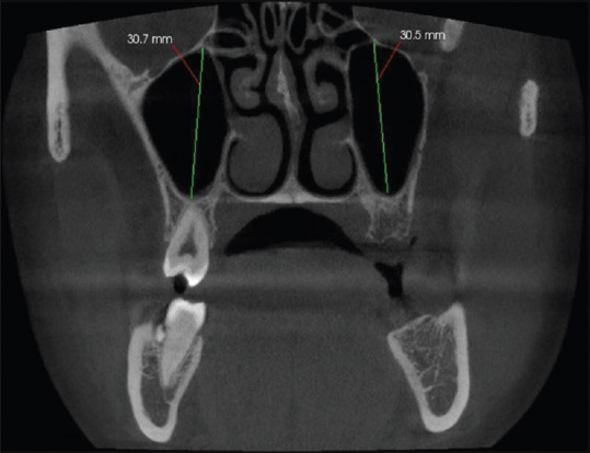
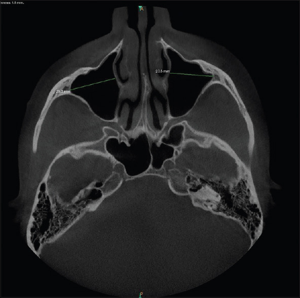
Materials and methods: The reporting of the study was done in accordance to strengthening the reporting of observational studies in epidemiology guidelines/checklist. Two hundred CBCT scans (400 MSs) were analysed for height and width of the maxillary sinuses (MSs) and its distribution with regards to gender. The prevalence of AMO and its different types of shapes were evaluated.
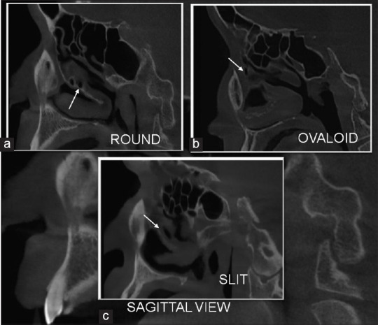
Results: Between the male and female groups, differences on both the right and left sides with respect to the MS height were observed (P > 0.05). However, the male group showed statistically significant higher values for right-sided MS width (P < 0.05). The prevalence of AMO in 400 MSs was found to be 65%, of which 63% of AMOs were present in the male group. The prevalence of types of accessory maxillary ostia revealed that the round and ovoid shapes were similar in number as compared to slit shapes.
Discussion: The forensic anthropology through CBCT can utilise the MS width as a tool to assess various anatomical variations. The frequency of AMOs can be utilised as a critical indicator for assessing such morphological and anatomical variations. As a result, the excellent spatial resolution of CBCT enables it as a reliable tool for identification of even smallest anatomical variations in MS.

 求助内容:
求助内容: 应助结果提醒方式:


