{"title":"原发性肋骨朗格汉斯细胞组织细胞增多症的临床特点及病理分析:病例分析及文献复习。","authors":"Guangsheng Ni, Yaxuan Ou, Runyu Ming, Jin Yang","doi":"10.21037/acr-24-251","DOIUrl":null,"url":null,"abstract":"<p><strong>Background: </strong>Langerhans cell histiocytosis (LCH) is a rare histiocytic neoplasm characterized by the abnormal proliferation of Langerhans cells, which can infiltrate various tissues throughout the body, leading to a spectrum of organ damage. This study aims to explore the clinical characteristics, imaging manifestations, and pathological features of the disease to enhance clinical diagnosis and understanding of such conditions.</p><p><strong>Case description: </strong>This study reviews the clinical data of four patients diagnosed with LCH at The First Affiliated Hospital of Hunan University of Traditional Chinese Medicine. All patients met the diagnostic criteria outlined in the LCH guidelines by the American Society of Hematology, with Langerhans cells expressing CD1a, S-100, and Langerin (CD207).</p><p><strong>Conclusions: </strong>LCH can involve various organs and systems, presenting with diverse clinical manifestations; in particular, rib Langerhans cell histiocytosis (RLCH) primarily manifests as asymptomatic or mildly painful bone swelling. Computerized tomography (CT) imaging of RLCH typically reveals localized masses and focal bone destruction, with or without surrounding soft tissue invasion. Conversely, magnetic resonance imaging provides a clearer assessment of lesion size and the extent of adjacent soft tissue involvement, offering advantages in guiding surgical excision. Diagnosis requires correlation with pathological and immunohistochemical results. For single-system, single-site (SS-s LCH), R0 surgical resection is feasible; however, single-system, multi-site (SS-m LCH) cases necessitate combined chemotherapy or targeted therapies. Overall, the treatment outcomes for this disease remain reasonably favorable.</p>","PeriodicalId":29752,"journal":{"name":"AME Case Reports","volume":"9 ","pages":"106"},"PeriodicalIF":0.7000,"publicationDate":"2025-07-15","publicationTypes":"Journal Article","fieldsOfStudy":null,"isOpenAccess":false,"openAccessPdf":"https://www.ncbi.nlm.nih.gov/pmc/articles/PMC12319616/pdf/","citationCount":"0","resultStr":"{\"title\":\"Clinical features and pathological analysis of primary rib Langerhans cell histiocytosis: case series and literature review.\",\"authors\":\"Guangsheng Ni, Yaxuan Ou, Runyu Ming, Jin Yang\",\"doi\":\"10.21037/acr-24-251\",\"DOIUrl\":null,\"url\":null,\"abstract\":\"<p><strong>Background: </strong>Langerhans cell histiocytosis (LCH) is a rare histiocytic neoplasm characterized by the abnormal proliferation of Langerhans cells, which can infiltrate various tissues throughout the body, leading to a spectrum of organ damage. This study aims to explore the clinical characteristics, imaging manifestations, and pathological features of the disease to enhance clinical diagnosis and understanding of such conditions.</p><p><strong>Case description: </strong>This study reviews the clinical data of four patients diagnosed with LCH at The First Affiliated Hospital of Hunan University of Traditional Chinese Medicine. All patients met the diagnostic criteria outlined in the LCH guidelines by the American Society of Hematology, with Langerhans cells expressing CD1a, S-100, and Langerin (CD207).</p><p><strong>Conclusions: </strong>LCH can involve various organs and systems, presenting with diverse clinical manifestations; in particular, rib Langerhans cell histiocytosis (RLCH) primarily manifests as asymptomatic or mildly painful bone swelling. Computerized tomography (CT) imaging of RLCH typically reveals localized masses and focal bone destruction, with or without surrounding soft tissue invasion. Conversely, magnetic resonance imaging provides a clearer assessment of lesion size and the extent of adjacent soft tissue involvement, offering advantages in guiding surgical excision. Diagnosis requires correlation with pathological and immunohistochemical results. For single-system, single-site (SS-s LCH), R0 surgical resection is feasible; however, single-system, multi-site (SS-m LCH) cases necessitate combined chemotherapy or targeted therapies. Overall, the treatment outcomes for this disease remain reasonably favorable.</p>\",\"PeriodicalId\":29752,\"journal\":{\"name\":\"AME Case Reports\",\"volume\":\"9 \",\"pages\":\"106\"},\"PeriodicalIF\":0.7000,\"publicationDate\":\"2025-07-15\",\"publicationTypes\":\"Journal Article\",\"fieldsOfStudy\":null,\"isOpenAccess\":false,\"openAccessPdf\":\"https://www.ncbi.nlm.nih.gov/pmc/articles/PMC12319616/pdf/\",\"citationCount\":\"0\",\"resultStr\":null,\"platform\":\"Semanticscholar\",\"paperid\":null,\"PeriodicalName\":\"AME Case Reports\",\"FirstCategoryId\":\"1085\",\"ListUrlMain\":\"https://doi.org/10.21037/acr-24-251\",\"RegionNum\":0,\"RegionCategory\":null,\"ArticlePicture\":[],\"TitleCN\":null,\"AbstractTextCN\":null,\"PMCID\":null,\"EPubDate\":\"2025/1/1 0:00:00\",\"PubModel\":\"eCollection\",\"JCR\":\"Q3\",\"JCRName\":\"MEDICINE, GENERAL & INTERNAL\",\"Score\":null,\"Total\":0}","platform":"Semanticscholar","paperid":null,"PeriodicalName":"AME Case Reports","FirstCategoryId":"1085","ListUrlMain":"https://doi.org/10.21037/acr-24-251","RegionNum":0,"RegionCategory":null,"ArticlePicture":[],"TitleCN":null,"AbstractTextCN":null,"PMCID":null,"EPubDate":"2025/1/1 0:00:00","PubModel":"eCollection","JCR":"Q3","JCRName":"MEDICINE, GENERAL & INTERNAL","Score":null,"Total":0}
Clinical features and pathological analysis of primary rib Langerhans cell histiocytosis: case series and literature review.
Background: Langerhans cell histiocytosis (LCH) is a rare histiocytic neoplasm characterized by the abnormal proliferation of Langerhans cells, which can infiltrate various tissues throughout the body, leading to a spectrum of organ damage. This study aims to explore the clinical characteristics, imaging manifestations, and pathological features of the disease to enhance clinical diagnosis and understanding of such conditions.
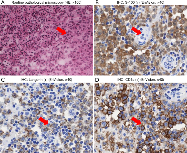
Case description: This study reviews the clinical data of four patients diagnosed with LCH at The First Affiliated Hospital of Hunan University of Traditional Chinese Medicine. All patients met the diagnostic criteria outlined in the LCH guidelines by the American Society of Hematology, with Langerhans cells expressing CD1a, S-100, and Langerin (CD207).
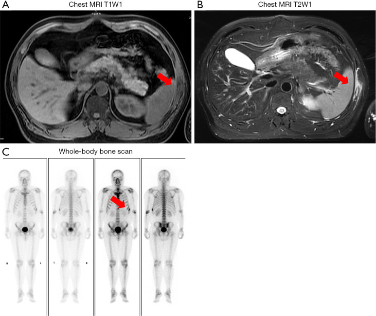
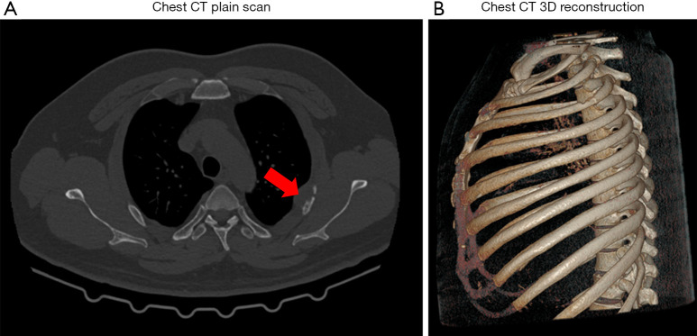
Conclusions: LCH can involve various organs and systems, presenting with diverse clinical manifestations; in particular, rib Langerhans cell histiocytosis (RLCH) primarily manifests as asymptomatic or mildly painful bone swelling. Computerized tomography (CT) imaging of RLCH typically reveals localized masses and focal bone destruction, with or without surrounding soft tissue invasion. Conversely, magnetic resonance imaging provides a clearer assessment of lesion size and the extent of adjacent soft tissue involvement, offering advantages in guiding surgical excision. Diagnosis requires correlation with pathological and immunohistochemical results. For single-system, single-site (SS-s LCH), R0 surgical resection is feasible; however, single-system, multi-site (SS-m LCH) cases necessitate combined chemotherapy or targeted therapies. Overall, the treatment outcomes for this disease remain reasonably favorable.

 求助内容:
求助内容: 应助结果提醒方式:


