Ana Cibrão-Pedroso, João Rocha-Neves, Rafael Vieira, João Barbosa Breda, André Ferreira
{"title":"颈动脉内膜切除术后视网膜和脉络膜厚度的变化:一项系统综述。","authors":"Ana Cibrão-Pedroso, João Rocha-Neves, Rafael Vieira, João Barbosa Breda, André Ferreira","doi":"10.1186/s40942-025-00713-1","DOIUrl":null,"url":null,"abstract":"<p><strong>Background: </strong>Carotid endarterectomy is a well-established procedure for enhancing cerebral perfusion in patients with internal carotid artery stenosis. As a multifactorial disease, carotid stenosis can have ocular implications, potentially affecting retinal and choroidal perfusion and contributing to visual dysfunction. This systematic review aims to evaluate changes in choroidal and retinal thickness after unilateral carotid endarterectomy, providing insight into the impact of the procedure on ocular perfusion.</p><p><strong>Methods: </strong>A comprehensive search was performed across PubMed, Scopus, and Web of Science up to July 2024, without restrictions on language or publication date. The inclusion criteria included original studies assessing retinal or choroidal thickness via optical coherence tomography before and after carotid endarterectomy in adults. Additional manual searches of reference lists and citation tracking were employed to ensure completeness. Study quality was appraised via the NHLBI tool for observational studies.</p><p><strong>Results: </strong>Six prospective observational studies involving 269 patients were included. Findings on choroidal thickness changes after carotid endarterectomy are heterogeneous. While two studies reported significant postoperative Choroidal Thickness increases-one within a week and another at three months-other studies reported no significant changes. One study suggested that higher degrees of carotid stenosis may blunt early Choroidal Thickness response. Retinal measurements were less consistently assessed; among the three studies that evaluated retinal nerve fibre layer and ganglion cell complex thickness, no consistent postoperative changes were observed. Overall, variability in study designs, Optical Coherence Tomography protocols, and follow-up durations limits comparability, precluding meta-analysis.</p><p><strong>Conclusions: </strong>This review highlights a potential association between carotid endarterectomy and improved ocular perfusion, as reflected by changes in choroidal thickness. However, inconsistencies across studies and limited data on retinal structural outcomes underscore the complexity of this relationship. These findings emphasize the need for larger, standardized studies to clarify the impact of carotid revascularization on the ocular microvasculature and guide future clinical practice.</p>","PeriodicalId":14289,"journal":{"name":"International Journal of Retina and Vitreous","volume":"11 1","pages":"91"},"PeriodicalIF":2.4000,"publicationDate":"2025-08-04","publicationTypes":"Journal Article","fieldsOfStudy":null,"isOpenAccess":false,"openAccessPdf":"https://www.ncbi.nlm.nih.gov/pmc/articles/PMC12323285/pdf/","citationCount":"0","resultStr":"{\"title\":\"Changes in retinal and choroidal thickness after carotid endarterectomy: a systematic review.\",\"authors\":\"Ana Cibrão-Pedroso, João Rocha-Neves, Rafael Vieira, João Barbosa Breda, André Ferreira\",\"doi\":\"10.1186/s40942-025-00713-1\",\"DOIUrl\":null,\"url\":null,\"abstract\":\"<p><strong>Background: </strong>Carotid endarterectomy is a well-established procedure for enhancing cerebral perfusion in patients with internal carotid artery stenosis. As a multifactorial disease, carotid stenosis can have ocular implications, potentially affecting retinal and choroidal perfusion and contributing to visual dysfunction. This systematic review aims to evaluate changes in choroidal and retinal thickness after unilateral carotid endarterectomy, providing insight into the impact of the procedure on ocular perfusion.</p><p><strong>Methods: </strong>A comprehensive search was performed across PubMed, Scopus, and Web of Science up to July 2024, without restrictions on language or publication date. The inclusion criteria included original studies assessing retinal or choroidal thickness via optical coherence tomography before and after carotid endarterectomy in adults. Additional manual searches of reference lists and citation tracking were employed to ensure completeness. Study quality was appraised via the NHLBI tool for observational studies.</p><p><strong>Results: </strong>Six prospective observational studies involving 269 patients were included. Findings on choroidal thickness changes after carotid endarterectomy are heterogeneous. While two studies reported significant postoperative Choroidal Thickness increases-one within a week and another at three months-other studies reported no significant changes. One study suggested that higher degrees of carotid stenosis may blunt early Choroidal Thickness response. Retinal measurements were less consistently assessed; among the three studies that evaluated retinal nerve fibre layer and ganglion cell complex thickness, no consistent postoperative changes were observed. Overall, variability in study designs, Optical Coherence Tomography protocols, and follow-up durations limits comparability, precluding meta-analysis.</p><p><strong>Conclusions: </strong>This review highlights a potential association between carotid endarterectomy and improved ocular perfusion, as reflected by changes in choroidal thickness. However, inconsistencies across studies and limited data on retinal structural outcomes underscore the complexity of this relationship. These findings emphasize the need for larger, standardized studies to clarify the impact of carotid revascularization on the ocular microvasculature and guide future clinical practice.</p>\",\"PeriodicalId\":14289,\"journal\":{\"name\":\"International Journal of Retina and Vitreous\",\"volume\":\"11 1\",\"pages\":\"91\"},\"PeriodicalIF\":2.4000,\"publicationDate\":\"2025-08-04\",\"publicationTypes\":\"Journal Article\",\"fieldsOfStudy\":null,\"isOpenAccess\":false,\"openAccessPdf\":\"https://www.ncbi.nlm.nih.gov/pmc/articles/PMC12323285/pdf/\",\"citationCount\":\"0\",\"resultStr\":null,\"platform\":\"Semanticscholar\",\"paperid\":null,\"PeriodicalName\":\"International Journal of Retina and Vitreous\",\"FirstCategoryId\":\"1085\",\"ListUrlMain\":\"https://doi.org/10.1186/s40942-025-00713-1\",\"RegionNum\":0,\"RegionCategory\":null,\"ArticlePicture\":[],\"TitleCN\":null,\"AbstractTextCN\":null,\"PMCID\":null,\"EPubDate\":\"\",\"PubModel\":\"\",\"JCR\":\"Q2\",\"JCRName\":\"OPHTHALMOLOGY\",\"Score\":null,\"Total\":0}","platform":"Semanticscholar","paperid":null,"PeriodicalName":"International Journal of Retina and Vitreous","FirstCategoryId":"1085","ListUrlMain":"https://doi.org/10.1186/s40942-025-00713-1","RegionNum":0,"RegionCategory":null,"ArticlePicture":[],"TitleCN":null,"AbstractTextCN":null,"PMCID":null,"EPubDate":"","PubModel":"","JCR":"Q2","JCRName":"OPHTHALMOLOGY","Score":null,"Total":0}
引用次数: 0
摘要
背景:颈动脉内膜切除术是颈内动脉狭窄患者增强脑灌注的一种成熟的手术方法。作为一种多因素疾病,颈动脉狭窄可影响眼部,潜在地影响视网膜和脉络膜灌注并导致视力障碍。本系统综述旨在评估单侧颈动脉内膜切除术后脉络膜和视网膜厚度的变化,为手术对眼灌注的影响提供见解。方法:综合检索PubMed、Scopus和Web of Science,截止到2024年7月,不受语言和出版日期的限制。纳入标准包括通过光学相干断层扫描评估成人颈动脉内膜切除术前后视网膜或脉络膜厚度的原始研究。额外的手动检索参考文献列表和引文跟踪,以确保完整性。通过观察性研究的NHLBI工具评价研究质量。结果:纳入了6项前瞻性观察性研究,涉及269例患者。颈动脉内膜切除术后脉络膜厚度变化的结果是不均匀的。虽然有两项研究报告术后脉络膜厚度显著增加——一项在一周内,另一项在三个月后——但其他研究报告没有显著变化。一项研究表明,颈动脉狭窄程度越高,早期脉络膜厚度反应越迟钝。视网膜测量的评估不太一致;在评估视网膜神经纤维层和神经节细胞复合体厚度的三项研究中,未观察到一致的术后变化。总的来说,研究设计、光学相干断层扫描方案和随访时间的可变性限制了可比性,排除了荟萃分析。结论:这篇综述强调了颈动脉内膜切除术与改善眼灌注之间的潜在关联,这反映在脉络膜厚度的改变上。然而,研究之间的不一致性和视网膜结构结果的有限数据强调了这种关系的复杂性。这些发现强调需要更大规模、标准化的研究来阐明颈动脉血管重建术对眼部微血管的影响,并指导未来的临床实践。
Changes in retinal and choroidal thickness after carotid endarterectomy: a systematic review.
Background: Carotid endarterectomy is a well-established procedure for enhancing cerebral perfusion in patients with internal carotid artery stenosis. As a multifactorial disease, carotid stenosis can have ocular implications, potentially affecting retinal and choroidal perfusion and contributing to visual dysfunction. This systematic review aims to evaluate changes in choroidal and retinal thickness after unilateral carotid endarterectomy, providing insight into the impact of the procedure on ocular perfusion.
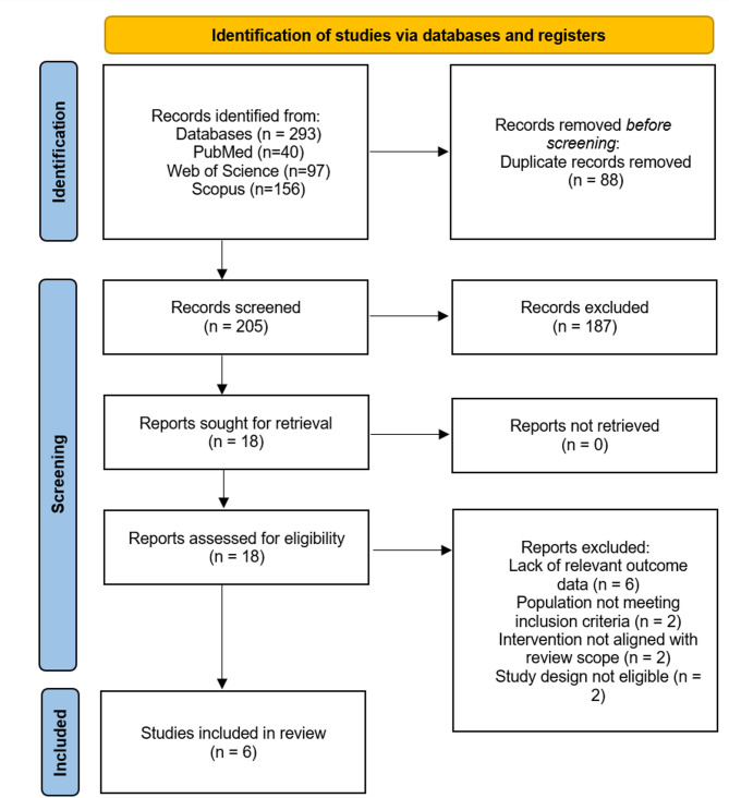
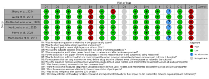
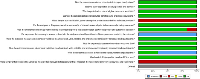
Methods: A comprehensive search was performed across PubMed, Scopus, and Web of Science up to July 2024, without restrictions on language or publication date. The inclusion criteria included original studies assessing retinal or choroidal thickness via optical coherence tomography before and after carotid endarterectomy in adults. Additional manual searches of reference lists and citation tracking were employed to ensure completeness. Study quality was appraised via the NHLBI tool for observational studies.
Results: Six prospective observational studies involving 269 patients were included. Findings on choroidal thickness changes after carotid endarterectomy are heterogeneous. While two studies reported significant postoperative Choroidal Thickness increases-one within a week and another at three months-other studies reported no significant changes. One study suggested that higher degrees of carotid stenosis may blunt early Choroidal Thickness response. Retinal measurements were less consistently assessed; among the three studies that evaluated retinal nerve fibre layer and ganglion cell complex thickness, no consistent postoperative changes were observed. Overall, variability in study designs, Optical Coherence Tomography protocols, and follow-up durations limits comparability, precluding meta-analysis.
Conclusions: This review highlights a potential association between carotid endarterectomy and improved ocular perfusion, as reflected by changes in choroidal thickness. However, inconsistencies across studies and limited data on retinal structural outcomes underscore the complexity of this relationship. These findings emphasize the need for larger, standardized studies to clarify the impact of carotid revascularization on the ocular microvasculature and guide future clinical practice.
期刊介绍:
International Journal of Retina and Vitreous focuses on the ophthalmic subspecialty of vitreoretinal disorders. The journal presents original articles on new approaches to diagnosis, outcomes of clinical trials, innovations in pharmacological therapy and surgical techniques, as well as basic science advances that impact clinical practice. Topical areas include, but are not limited to: -Imaging of the retina, choroid and vitreous -Innovations in optical coherence tomography (OCT) -Small-gauge vitrectomy, retinal detachment, chromovitrectomy -Electroretinography (ERG), microperimetry, other functional tests -Intraocular tumors -Retinal pharmacotherapy & drug delivery -Diabetic retinopathy & other vascular diseases -Age-related macular degeneration (AMD) & other macular entities

 求助内容:
求助内容: 应助结果提醒方式:


