Yue Hu, Xin Cao, Hongyi Chen, Daoying Geng, Kun Lv
{"title":"全脑静息状态fMRI特征的机器学习用于额叶胶质瘤的个体化分级。","authors":"Yue Hu, Xin Cao, Hongyi Chen, Daoying Geng, Kun Lv","doi":"10.1186/s40644-025-00920-x","DOIUrl":null,"url":null,"abstract":"<p><strong>Purpose: </strong>Accurate preoperative grading of gliomas is critical for therapeutic planning and prognostic evaluation. We developed a noninvasive machine learning model leveraging whole-brain resting-state functional magnetic resonance imaging (rs-fMRI) biomarkers to discriminate high-grade (HGGs) and low-grade gliomas (LGGs) in the frontal lobe.</p><p><strong>Methods: </strong>This retrospective study included 138 patients (78 LGGs, 60 HGGs) with left frontal gliomas. A total of 7134 features were extracted from the mean amplitude of low-frequency fluctuation (mALFF), mean fractional ALFF, mean percentage amplitude of fluctuation (mPerAF), mean regional homogeneity (mReHo) maps and resting-state functional connectivity (RSFC) matrix. Twelve predictive features were selected through Mann-Whitney U test, correlation analysis and least absolute shrinkage and selection operator method. The patients were stratified and randomized into the training and testing datasets with a 7:3 ratio. The logical regression, random forest, support vector machine (SVM) and adaptive boosting algorithms were used to establish models. The model performance was evaluated using area under the receiver operating characteristic curve, accuracy, sensitivity, and specificity.</p><p><strong>Results: </strong>The selected 12 features included 7 RSFC features, 4 mPerAF features, and 1 mReHo feature. Based on these features, the model was established using the SVM had an optimal performance. The accuracy in the training and testing datasets was 0.957 and 0.727, respectively. The area under the receiver operating characteristic curves was 0.972 and 0.799, respectively.</p><p><strong>Conclusions: </strong>Our whole-brain rs-fMRI radiomics approach provides an objective tool for preoperative glioma stratification. The biological interpretability of selected features reflects distinct neuroplasticity patterns between LGGs and HGGs, advancing understanding of glioma-network interactions.</p>","PeriodicalId":9548,"journal":{"name":"Cancer Imaging","volume":"25 1","pages":"97"},"PeriodicalIF":3.5000,"publicationDate":"2025-08-04","publicationTypes":"Journal Article","fieldsOfStudy":null,"isOpenAccess":false,"openAccessPdf":"https://www.ncbi.nlm.nih.gov/pmc/articles/PMC12323070/pdf/","citationCount":"0","resultStr":"{\"title\":\"Machine learning of whole-brain resting-state fMRI signatures for individualized grading of frontal gliomas.\",\"authors\":\"Yue Hu, Xin Cao, Hongyi Chen, Daoying Geng, Kun Lv\",\"doi\":\"10.1186/s40644-025-00920-x\",\"DOIUrl\":null,\"url\":null,\"abstract\":\"<p><strong>Purpose: </strong>Accurate preoperative grading of gliomas is critical for therapeutic planning and prognostic evaluation. We developed a noninvasive machine learning model leveraging whole-brain resting-state functional magnetic resonance imaging (rs-fMRI) biomarkers to discriminate high-grade (HGGs) and low-grade gliomas (LGGs) in the frontal lobe.</p><p><strong>Methods: </strong>This retrospective study included 138 patients (78 LGGs, 60 HGGs) with left frontal gliomas. A total of 7134 features were extracted from the mean amplitude of low-frequency fluctuation (mALFF), mean fractional ALFF, mean percentage amplitude of fluctuation (mPerAF), mean regional homogeneity (mReHo) maps and resting-state functional connectivity (RSFC) matrix. Twelve predictive features were selected through Mann-Whitney U test, correlation analysis and least absolute shrinkage and selection operator method. The patients were stratified and randomized into the training and testing datasets with a 7:3 ratio. The logical regression, random forest, support vector machine (SVM) and adaptive boosting algorithms were used to establish models. The model performance was evaluated using area under the receiver operating characteristic curve, accuracy, sensitivity, and specificity.</p><p><strong>Results: </strong>The selected 12 features included 7 RSFC features, 4 mPerAF features, and 1 mReHo feature. Based on these features, the model was established using the SVM had an optimal performance. The accuracy in the training and testing datasets was 0.957 and 0.727, respectively. The area under the receiver operating characteristic curves was 0.972 and 0.799, respectively.</p><p><strong>Conclusions: </strong>Our whole-brain rs-fMRI radiomics approach provides an objective tool for preoperative glioma stratification. The biological interpretability of selected features reflects distinct neuroplasticity patterns between LGGs and HGGs, advancing understanding of glioma-network interactions.</p>\",\"PeriodicalId\":9548,\"journal\":{\"name\":\"Cancer Imaging\",\"volume\":\"25 1\",\"pages\":\"97\"},\"PeriodicalIF\":3.5000,\"publicationDate\":\"2025-08-04\",\"publicationTypes\":\"Journal Article\",\"fieldsOfStudy\":null,\"isOpenAccess\":false,\"openAccessPdf\":\"https://www.ncbi.nlm.nih.gov/pmc/articles/PMC12323070/pdf/\",\"citationCount\":\"0\",\"resultStr\":null,\"platform\":\"Semanticscholar\",\"paperid\":null,\"PeriodicalName\":\"Cancer Imaging\",\"FirstCategoryId\":\"3\",\"ListUrlMain\":\"https://doi.org/10.1186/s40644-025-00920-x\",\"RegionNum\":2,\"RegionCategory\":\"医学\",\"ArticlePicture\":[],\"TitleCN\":null,\"AbstractTextCN\":null,\"PMCID\":null,\"EPubDate\":\"\",\"PubModel\":\"\",\"JCR\":\"Q2\",\"JCRName\":\"ONCOLOGY\",\"Score\":null,\"Total\":0}","platform":"Semanticscholar","paperid":null,"PeriodicalName":"Cancer Imaging","FirstCategoryId":"3","ListUrlMain":"https://doi.org/10.1186/s40644-025-00920-x","RegionNum":2,"RegionCategory":"医学","ArticlePicture":[],"TitleCN":null,"AbstractTextCN":null,"PMCID":null,"EPubDate":"","PubModel":"","JCR":"Q2","JCRName":"ONCOLOGY","Score":null,"Total":0}
Machine learning of whole-brain resting-state fMRI signatures for individualized grading of frontal gliomas.
Purpose: Accurate preoperative grading of gliomas is critical for therapeutic planning and prognostic evaluation. We developed a noninvasive machine learning model leveraging whole-brain resting-state functional magnetic resonance imaging (rs-fMRI) biomarkers to discriminate high-grade (HGGs) and low-grade gliomas (LGGs) in the frontal lobe.
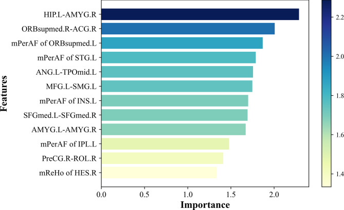
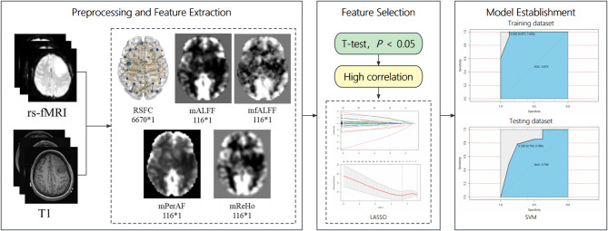
Methods: This retrospective study included 138 patients (78 LGGs, 60 HGGs) with left frontal gliomas. A total of 7134 features were extracted from the mean amplitude of low-frequency fluctuation (mALFF), mean fractional ALFF, mean percentage amplitude of fluctuation (mPerAF), mean regional homogeneity (mReHo) maps and resting-state functional connectivity (RSFC) matrix. Twelve predictive features were selected through Mann-Whitney U test, correlation analysis and least absolute shrinkage and selection operator method. The patients were stratified and randomized into the training and testing datasets with a 7:3 ratio. The logical regression, random forest, support vector machine (SVM) and adaptive boosting algorithms were used to establish models. The model performance was evaluated using area under the receiver operating characteristic curve, accuracy, sensitivity, and specificity.
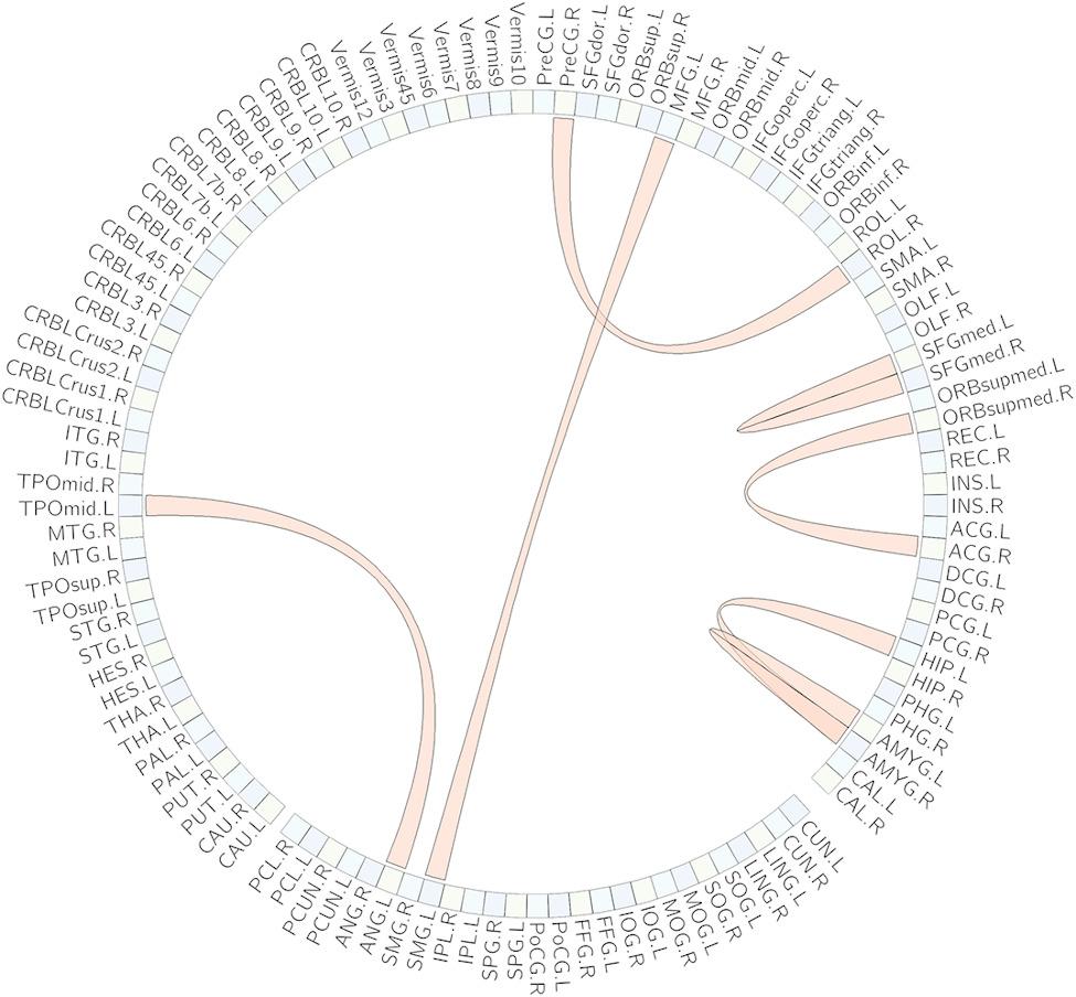
Results: The selected 12 features included 7 RSFC features, 4 mPerAF features, and 1 mReHo feature. Based on these features, the model was established using the SVM had an optimal performance. The accuracy in the training and testing datasets was 0.957 and 0.727, respectively. The area under the receiver operating characteristic curves was 0.972 and 0.799, respectively.
Conclusions: Our whole-brain rs-fMRI radiomics approach provides an objective tool for preoperative glioma stratification. The biological interpretability of selected features reflects distinct neuroplasticity patterns between LGGs and HGGs, advancing understanding of glioma-network interactions.
Cancer ImagingONCOLOGY-RADIOLOGY, NUCLEAR MEDICINE & MEDICAL IMAGING
CiteScore
7.00
自引率
0.00%
发文量
66
审稿时长
>12 weeks
期刊介绍:
Cancer Imaging is an open access, peer-reviewed journal publishing original articles, reviews and editorials written by expert international radiologists working in oncology.
The journal encompasses CT, MR, PET, ultrasound, radionuclide and multimodal imaging in all kinds of malignant tumours, plus new developments, techniques and innovations. Topics of interest include:
Breast Imaging
Chest
Complications of treatment
Ear, Nose & Throat
Gastrointestinal
Hepatobiliary & Pancreatic
Imaging biomarkers
Interventional
Lymphoma
Measurement of tumour response
Molecular functional imaging
Musculoskeletal
Neuro oncology
Nuclear Medicine
Paediatric.

 求助内容:
求助内容: 应助结果提醒方式:


