C F Agudelo, B Lukac, A Bockay, V Vargova, G Kackova, M Figurova, S Hornak
{"title":"犬偶发左冠状动脉至肺动脉主动脉瘘1例。","authors":"C F Agudelo, B Lukac, A Bockay, V Vargova, G Kackova, M Figurova, S Hornak","doi":"10.1186/s12917-025-04730-y","DOIUrl":null,"url":null,"abstract":"<p><p>A vascular communication between the left coronary artery and the main pulmonary artery (coronary fistula) was incidentally diagnosed in a 3-year-old female Tatran hound during a pre-anesthetic evaluation using echocardiography. Trauma and endocarditis were not suspected given the patient's history, clinical examination, and diagnostic imaging, which could have contributed to the development of this condition. Transthoracic echocardiography revealed dilation of the left coronary ostium, and continuous shunting on color and spectral Doppler was observed and this led to the diagnosis of coronary artery fistula. This diagnosis was confirmed through computed tomography. Coronary artery fistulas are infrequent findings in both humans and animals. According to the authors, this is the first reported clinical case of a congenital fistula between the left coronary artery and the main pulmonary artery in a dog.</p>","PeriodicalId":9041,"journal":{"name":"BMC Veterinary Research","volume":"21 1","pages":"503"},"PeriodicalIF":2.6000,"publicationDate":"2025-08-04","publicationTypes":"Journal Article","fieldsOfStudy":null,"isOpenAccess":false,"openAccessPdf":"https://www.ncbi.nlm.nih.gov/pmc/articles/PMC12323072/pdf/","citationCount":"0","resultStr":"{\"title\":\"Case report of an incidental left coronary artery to main pulmonary artery fistula in a dog.\",\"authors\":\"C F Agudelo, B Lukac, A Bockay, V Vargova, G Kackova, M Figurova, S Hornak\",\"doi\":\"10.1186/s12917-025-04730-y\",\"DOIUrl\":null,\"url\":null,\"abstract\":\"<p><p>A vascular communication between the left coronary artery and the main pulmonary artery (coronary fistula) was incidentally diagnosed in a 3-year-old female Tatran hound during a pre-anesthetic evaluation using echocardiography. Trauma and endocarditis were not suspected given the patient's history, clinical examination, and diagnostic imaging, which could have contributed to the development of this condition. Transthoracic echocardiography revealed dilation of the left coronary ostium, and continuous shunting on color and spectral Doppler was observed and this led to the diagnosis of coronary artery fistula. This diagnosis was confirmed through computed tomography. Coronary artery fistulas are infrequent findings in both humans and animals. According to the authors, this is the first reported clinical case of a congenital fistula between the left coronary artery and the main pulmonary artery in a dog.</p>\",\"PeriodicalId\":9041,\"journal\":{\"name\":\"BMC Veterinary Research\",\"volume\":\"21 1\",\"pages\":\"503\"},\"PeriodicalIF\":2.6000,\"publicationDate\":\"2025-08-04\",\"publicationTypes\":\"Journal Article\",\"fieldsOfStudy\":null,\"isOpenAccess\":false,\"openAccessPdf\":\"https://www.ncbi.nlm.nih.gov/pmc/articles/PMC12323072/pdf/\",\"citationCount\":\"0\",\"resultStr\":null,\"platform\":\"Semanticscholar\",\"paperid\":null,\"PeriodicalName\":\"BMC Veterinary Research\",\"FirstCategoryId\":\"97\",\"ListUrlMain\":\"https://doi.org/10.1186/s12917-025-04730-y\",\"RegionNum\":2,\"RegionCategory\":\"农林科学\",\"ArticlePicture\":[],\"TitleCN\":null,\"AbstractTextCN\":null,\"PMCID\":null,\"EPubDate\":\"\",\"PubModel\":\"\",\"JCR\":\"Q1\",\"JCRName\":\"VETERINARY SCIENCES\",\"Score\":null,\"Total\":0}","platform":"Semanticscholar","paperid":null,"PeriodicalName":"BMC Veterinary Research","FirstCategoryId":"97","ListUrlMain":"https://doi.org/10.1186/s12917-025-04730-y","RegionNum":2,"RegionCategory":"农林科学","ArticlePicture":[],"TitleCN":null,"AbstractTextCN":null,"PMCID":null,"EPubDate":"","PubModel":"","JCR":"Q1","JCRName":"VETERINARY SCIENCES","Score":null,"Total":0}
Case report of an incidental left coronary artery to main pulmonary artery fistula in a dog.
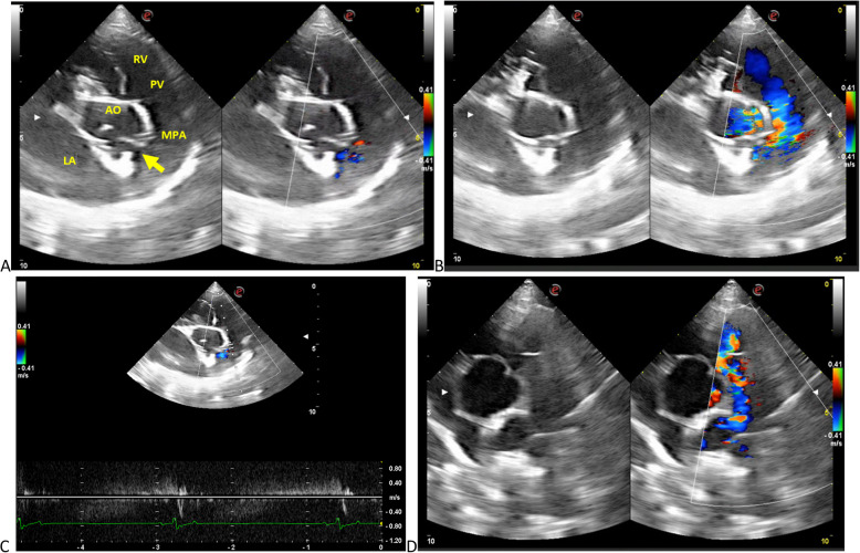
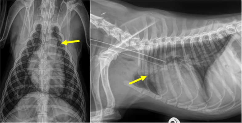
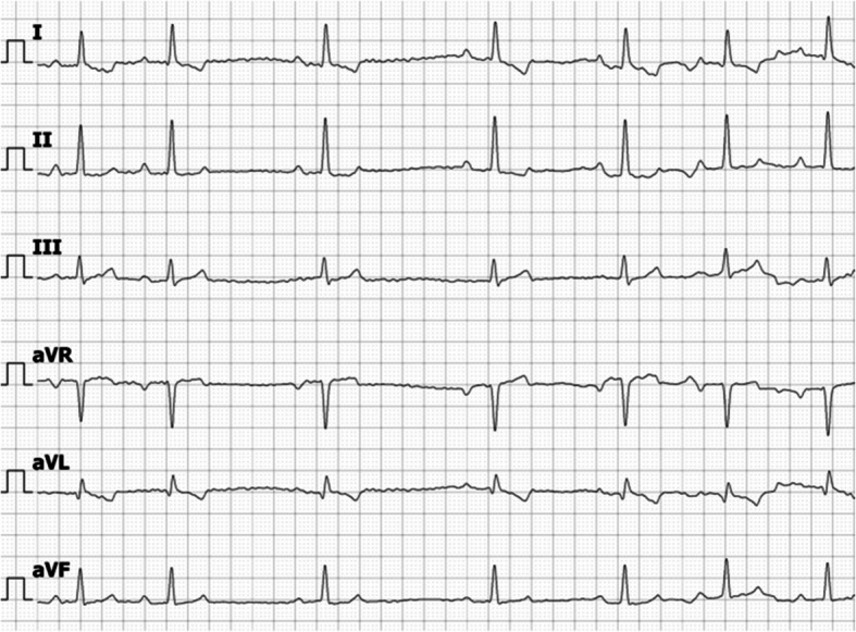
A vascular communication between the left coronary artery and the main pulmonary artery (coronary fistula) was incidentally diagnosed in a 3-year-old female Tatran hound during a pre-anesthetic evaluation using echocardiography. Trauma and endocarditis were not suspected given the patient's history, clinical examination, and diagnostic imaging, which could have contributed to the development of this condition. Transthoracic echocardiography revealed dilation of the left coronary ostium, and continuous shunting on color and spectral Doppler was observed and this led to the diagnosis of coronary artery fistula. This diagnosis was confirmed through computed tomography. Coronary artery fistulas are infrequent findings in both humans and animals. According to the authors, this is the first reported clinical case of a congenital fistula between the left coronary artery and the main pulmonary artery in a dog.
期刊介绍:
BMC Veterinary Research is an open access, peer-reviewed journal that considers articles on all aspects of veterinary science and medicine, including the epidemiology, diagnosis, prevention and treatment of medical conditions of domestic, companion, farm and wild animals, as well as the biomedical processes that underlie their health.

 求助内容:
求助内容: 应助结果提醒方式:


