{"title":"马颅内肿瘤的诊断病理学","authors":"H. G. Miller, A. Suarez-Bonnet","doi":"10.1111/eve.14145","DOIUrl":null,"url":null,"abstract":"<p>Neoplasia is rare in horses and central nervous system (CNS) neoplasia is so rare that, pituitary adenomas excluded, no specific intracranial neoplasms are mentioned in Cotchin's preliminary work describing the epidemiology of equine neoplasia (Cotchin, <span>1977</span>). Moreover, an early survey identified 28 nervous tissue tumours in horses but none were reported in the central nervous system (Hayes Jr. et al., <span>1975</span>). However, as de Sousa et al. (<span>2025</span>) highlight in the accompanying case report, equine intracranial neoplasia can cause significant clinical disease and histopathology is essential to making a complete diagnosis, albeit inevitably through post-mortem examination. Herein follows a brief discussion of a variety of intracranial tumours reported in horses and their histopathological diagnostic features with a summary of their immunohistochemical (IHC) characteristics in Table 1. Non-neoplastic causes of intracranial space-occupying lesions, for example, cholesterol granulomas, must always remain on the list of differential diagnoses but lie outside the remit of this commentary. Any readers seeking further details and descriptions of the characteristics of these lesions are encouraged to pursue some of the references at the end of this commentary, or indeed the associated case report.</p><p>There are a variety of reported intracranial neoplasms despite them remaining a rare diagnosis that relies on post-mortem examination. While the majority of types of tumours only exert clinical effects through a local mass effect, acting as benign space-occupying lesions, some neoplasms are malignant and the most common presentation of intracranial neoplasia remains PPID. The authors hope readers can better appreciate the heterogeneity of intracranial neoplasia and take home a greater understanding of how histological patterns allow pathologists to distinguish between grossly very similar masses.</p><p><b>H. G. Miller:</b> Conceptualization; writing – original draft; writing – review and editing. <b>A. Suarez-Bonnet:</b> Conceptualization; writing – original draft; writing – review and editing.</p><p>There are no funders to report for this submission.</p><p>No conflicts of interest have been declared.</p><p>Not required for this clinical commentary.</p>","PeriodicalId":11786,"journal":{"name":"Equine Veterinary Education","volume":"37 9","pages":"456-460"},"PeriodicalIF":0.8000,"publicationDate":"2025-03-24","publicationTypes":"Journal Article","fieldsOfStudy":null,"isOpenAccess":false,"openAccessPdf":"https://onlinelibrary.wiley.com/doi/epdf/10.1111/eve.14145","citationCount":"0","resultStr":"{\"title\":\"Diagnostic pathology of equine intracranial neoplasms\",\"authors\":\"H. G. Miller, A. Suarez-Bonnet\",\"doi\":\"10.1111/eve.14145\",\"DOIUrl\":null,\"url\":null,\"abstract\":\"<p>Neoplasia is rare in horses and central nervous system (CNS) neoplasia is so rare that, pituitary adenomas excluded, no specific intracranial neoplasms are mentioned in Cotchin's preliminary work describing the epidemiology of equine neoplasia (Cotchin, <span>1977</span>). Moreover, an early survey identified 28 nervous tissue tumours in horses but none were reported in the central nervous system (Hayes Jr. et al., <span>1975</span>). However, as de Sousa et al. (<span>2025</span>) highlight in the accompanying case report, equine intracranial neoplasia can cause significant clinical disease and histopathology is essential to making a complete diagnosis, albeit inevitably through post-mortem examination. Herein follows a brief discussion of a variety of intracranial tumours reported in horses and their histopathological diagnostic features with a summary of their immunohistochemical (IHC) characteristics in Table 1. Non-neoplastic causes of intracranial space-occupying lesions, for example, cholesterol granulomas, must always remain on the list of differential diagnoses but lie outside the remit of this commentary. Any readers seeking further details and descriptions of the characteristics of these lesions are encouraged to pursue some of the references at the end of this commentary, or indeed the associated case report.</p><p>There are a variety of reported intracranial neoplasms despite them remaining a rare diagnosis that relies on post-mortem examination. While the majority of types of tumours only exert clinical effects through a local mass effect, acting as benign space-occupying lesions, some neoplasms are malignant and the most common presentation of intracranial neoplasia remains PPID. The authors hope readers can better appreciate the heterogeneity of intracranial neoplasia and take home a greater understanding of how histological patterns allow pathologists to distinguish between grossly very similar masses.</p><p><b>H. G. Miller:</b> Conceptualization; writing – original draft; writing – review and editing. <b>A. Suarez-Bonnet:</b> Conceptualization; writing – original draft; writing – review and editing.</p><p>There are no funders to report for this submission.</p><p>No conflicts of interest have been declared.</p><p>Not required for this clinical commentary.</p>\",\"PeriodicalId\":11786,\"journal\":{\"name\":\"Equine Veterinary Education\",\"volume\":\"37 9\",\"pages\":\"456-460\"},\"PeriodicalIF\":0.8000,\"publicationDate\":\"2025-03-24\",\"publicationTypes\":\"Journal Article\",\"fieldsOfStudy\":null,\"isOpenAccess\":false,\"openAccessPdf\":\"https://onlinelibrary.wiley.com/doi/epdf/10.1111/eve.14145\",\"citationCount\":\"0\",\"resultStr\":null,\"platform\":\"Semanticscholar\",\"paperid\":null,\"PeriodicalName\":\"Equine Veterinary Education\",\"FirstCategoryId\":\"97\",\"ListUrlMain\":\"https://beva.onlinelibrary.wiley.com/doi/10.1111/eve.14145\",\"RegionNum\":4,\"RegionCategory\":\"农林科学\",\"ArticlePicture\":[],\"TitleCN\":null,\"AbstractTextCN\":null,\"PMCID\":null,\"EPubDate\":\"\",\"PubModel\":\"\",\"JCR\":\"Q3\",\"JCRName\":\"VETERINARY SCIENCES\",\"Score\":null,\"Total\":0}","platform":"Semanticscholar","paperid":null,"PeriodicalName":"Equine Veterinary Education","FirstCategoryId":"97","ListUrlMain":"https://beva.onlinelibrary.wiley.com/doi/10.1111/eve.14145","RegionNum":4,"RegionCategory":"农林科学","ArticlePicture":[],"TitleCN":null,"AbstractTextCN":null,"PMCID":null,"EPubDate":"","PubModel":"","JCR":"Q3","JCRName":"VETERINARY SCIENCES","Score":null,"Total":0}
引用次数: 0
摘要
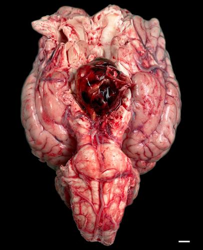
瘤变在马中很少见,而且中枢神经系统(CNS)瘤变非常罕见,在Cotchin描述马瘤变流行病学的初步工作中(Cotchin, 1977),除垂体腺瘤外,没有提到特异性颅内肿瘤。此外,一项早期调查在马身上发现了28种神经组织肿瘤,但没有在中枢神经系统中发现(Hayes Jr. et al., 1975)。然而,正如de Sousa等人(2025)在附带的病例报告中强调的那样,马颅内肿瘤可引起重大的临床疾病,组织病理学对于做出完整的诊断至关重要,尽管不可避免地要通过死后检查。本文简要讨论了在马中报道的各种颅内肿瘤及其组织病理学诊断特征,并在表1中总结了其免疫组织化学(IHC)特征。颅内占位性病变的非肿瘤性原因,例如胆固醇肉芽肿,必须始终保留在鉴别诊断列表中,但不在本评论的范围内。任何寻求这些病变特征的进一步细节和描述的读者都被鼓励去追求一些参考文献在这篇评论的末尾,或者确实相关的病例报告。有各种各样的颅内肿瘤报道,尽管他们仍然是一个罕见的诊断,依赖于尸检。虽然大多数类型的肿瘤仅通过局部肿块效应发挥临床作用,作为良性占位性病变,但一些肿瘤是恶性的,颅内肿瘤最常见的表现仍然是PPID。作者希望读者能够更好地认识颅内肿瘤的异质性,并对病理学家如何区分非常相似的肿块有更深入的了解。G. Miller:概念化;写作——原稿;写作——审阅和编辑。A.苏亚雷斯-邦纳:概念化;写作——原稿;写作——审阅和编辑。本次提交无需报告资助者。没有宣布利益冲突。这篇临床评论不需要。
Diagnostic pathology of equine intracranial neoplasms
Neoplasia is rare in horses and central nervous system (CNS) neoplasia is so rare that, pituitary adenomas excluded, no specific intracranial neoplasms are mentioned in Cotchin's preliminary work describing the epidemiology of equine neoplasia (Cotchin, 1977). Moreover, an early survey identified 28 nervous tissue tumours in horses but none were reported in the central nervous system (Hayes Jr. et al., 1975). However, as de Sousa et al. (2025) highlight in the accompanying case report, equine intracranial neoplasia can cause significant clinical disease and histopathology is essential to making a complete diagnosis, albeit inevitably through post-mortem examination. Herein follows a brief discussion of a variety of intracranial tumours reported in horses and their histopathological diagnostic features with a summary of their immunohistochemical (IHC) characteristics in Table 1. Non-neoplastic causes of intracranial space-occupying lesions, for example, cholesterol granulomas, must always remain on the list of differential diagnoses but lie outside the remit of this commentary. Any readers seeking further details and descriptions of the characteristics of these lesions are encouraged to pursue some of the references at the end of this commentary, or indeed the associated case report.
There are a variety of reported intracranial neoplasms despite them remaining a rare diagnosis that relies on post-mortem examination. While the majority of types of tumours only exert clinical effects through a local mass effect, acting as benign space-occupying lesions, some neoplasms are malignant and the most common presentation of intracranial neoplasia remains PPID. The authors hope readers can better appreciate the heterogeneity of intracranial neoplasia and take home a greater understanding of how histological patterns allow pathologists to distinguish between grossly very similar masses.
H. G. Miller: Conceptualization; writing – original draft; writing – review and editing. A. Suarez-Bonnet: Conceptualization; writing – original draft; writing – review and editing.
There are no funders to report for this submission.
期刊介绍:
Equine Veterinary Education (EVE) is the official journal of post-graduate education of both the British Equine Veterinary Association (BEVA) and the American Association of Equine Practitioners (AAEP).
Equine Veterinary Education is a monthly, peer-reviewed, subscription-based journal, integrating clinical research papers, review articles and case reports from international sources, covering all aspects of medicine and surgery relating to equids. These papers facilitate the dissemination and implementation of new ideas and techniques relating to clinical veterinary practice, with the ultimate aim of promoting best practice. New developments are placed in perspective, encompassing new concepts and peer commentary. The target audience is veterinarians primarily engaged in the practise of equine medicine and surgery. The educational value of a submitted article is one of the most important criteria that are assessed when deciding whether to accept it for publication. Articles do not necessarily need to contain original or novel information but we welcome submission of this material. The educational value of an article may relate to articles published with it (e.g. a Case Report may not have direct educational value but an associated Clinical Commentary or Review Article published alongside it will enhance the educational value).


 求助内容:
求助内容: 应助结果提醒方式:


