Fernanda Barthelson Carvalho de Moura, Victor Gustavo Santos Môra, Natalia Camargo Faraldo, Gabriel Correa de Camargo, Carlos Roberto Teixeira, Maria Valeria de Toledo Rodovalho, Tatiane Terumi Negrão Watanabe, Daniel Felipe Barrantes Murillo, Noeme Sousa Rocha, Carlos Eduardo Fonseca-Alves
{"title":"散养大食蚁兽阴茎的形态、组织化学和免疫组织化学特征","authors":"Fernanda Barthelson Carvalho de Moura, Victor Gustavo Santos Môra, Natalia Camargo Faraldo, Gabriel Correa de Camargo, Carlos Roberto Teixeira, Maria Valeria de Toledo Rodovalho, Tatiane Terumi Negrão Watanabe, Daniel Felipe Barrantes Murillo, Noeme Sousa Rocha, Carlos Eduardo Fonseca-Alves","doi":"10.1111/ahe.70057","DOIUrl":null,"url":null,"abstract":"<p>The giant anteater (<i>Myrmecophaga tridactyla</i>) belongs to the superorder Xenarthra and is distributed throughout Central and South America. This animal is listed as ‘vulnerable’ on the International Union for Conservation of Nature's Red List of Threatened Species. Studies on the reproduction of this species are essential because of its peculiarities; however, there is a lack of information on its reproductive aspects and the biotechnologies that can be applied to it. Morphological and histopathological descriptions of the reproductive organs of <i>Myrmecophaga tridactyla</i> are fundamental for determining the general characteristics that could improve our understanding of reproductive disorders in this species. Therefore, this study aimed to perform morphological and histochemical characterisation of the penis of <i>Myrmecophaga tridactyla</i>. For this purpose, we collected six postmortem samples of giant anteater penises. The penis of the giant anteater has no foreskin, two erectile bodies, a paired corpora cavernosa, corpus spongiosum and urethra. Type I (red) and type III (yellow-green) collagen fibres were distributed throughout the stroma and erectile bodies of the penis. Periodic acid-Schiff (PAS)-positive staining was observed in the epithelial cells at the base of the hair follicles, and immunohistochemical immunolabelling for androgen receptors (AR) and oestrogen receptors (ER) was detected in all cells of the transitional epithelium of the penile urethra. These findings suggest that penile urethra cells are sensitive to oestrogen and progesterone.</p>","PeriodicalId":49290,"journal":{"name":"Anatomia Histologia Embryologia","volume":"54 4","pages":""},"PeriodicalIF":1.0000,"publicationDate":"2025-08-06","publicationTypes":"Journal Article","fieldsOfStudy":null,"isOpenAccess":false,"openAccessPdf":"https://onlinelibrary.wiley.com/doi/epdf/10.1111/ahe.70057","citationCount":"0","resultStr":"{\"title\":\"Morphological, Histochemical and Immunohistochemical Characterisation of the Penis in Free-Ranging Giant Anteaters (Myrmecophaga tridactyla)\",\"authors\":\"Fernanda Barthelson Carvalho de Moura, Victor Gustavo Santos Môra, Natalia Camargo Faraldo, Gabriel Correa de Camargo, Carlos Roberto Teixeira, Maria Valeria de Toledo Rodovalho, Tatiane Terumi Negrão Watanabe, Daniel Felipe Barrantes Murillo, Noeme Sousa Rocha, Carlos Eduardo Fonseca-Alves\",\"doi\":\"10.1111/ahe.70057\",\"DOIUrl\":null,\"url\":null,\"abstract\":\"<p>The giant anteater (<i>Myrmecophaga tridactyla</i>) belongs to the superorder Xenarthra and is distributed throughout Central and South America. This animal is listed as ‘vulnerable’ on the International Union for Conservation of Nature's Red List of Threatened Species. Studies on the reproduction of this species are essential because of its peculiarities; however, there is a lack of information on its reproductive aspects and the biotechnologies that can be applied to it. Morphological and histopathological descriptions of the reproductive organs of <i>Myrmecophaga tridactyla</i> are fundamental for determining the general characteristics that could improve our understanding of reproductive disorders in this species. Therefore, this study aimed to perform morphological and histochemical characterisation of the penis of <i>Myrmecophaga tridactyla</i>. For this purpose, we collected six postmortem samples of giant anteater penises. The penis of the giant anteater has no foreskin, two erectile bodies, a paired corpora cavernosa, corpus spongiosum and urethra. Type I (red) and type III (yellow-green) collagen fibres were distributed throughout the stroma and erectile bodies of the penis. Periodic acid-Schiff (PAS)-positive staining was observed in the epithelial cells at the base of the hair follicles, and immunohistochemical immunolabelling for androgen receptors (AR) and oestrogen receptors (ER) was detected in all cells of the transitional epithelium of the penile urethra. These findings suggest that penile urethra cells are sensitive to oestrogen and progesterone.</p>\",\"PeriodicalId\":49290,\"journal\":{\"name\":\"Anatomia Histologia Embryologia\",\"volume\":\"54 4\",\"pages\":\"\"},\"PeriodicalIF\":1.0000,\"publicationDate\":\"2025-08-06\",\"publicationTypes\":\"Journal Article\",\"fieldsOfStudy\":null,\"isOpenAccess\":false,\"openAccessPdf\":\"https://onlinelibrary.wiley.com/doi/epdf/10.1111/ahe.70057\",\"citationCount\":\"0\",\"resultStr\":null,\"platform\":\"Semanticscholar\",\"paperid\":null,\"PeriodicalName\":\"Anatomia Histologia Embryologia\",\"FirstCategoryId\":\"97\",\"ListUrlMain\":\"https://onlinelibrary.wiley.com/doi/10.1111/ahe.70057\",\"RegionNum\":4,\"RegionCategory\":\"农林科学\",\"ArticlePicture\":[],\"TitleCN\":null,\"AbstractTextCN\":null,\"PMCID\":null,\"EPubDate\":\"\",\"PubModel\":\"\",\"JCR\":\"Q4\",\"JCRName\":\"ANATOMY & MORPHOLOGY\",\"Score\":null,\"Total\":0}","platform":"Semanticscholar","paperid":null,"PeriodicalName":"Anatomia Histologia Embryologia","FirstCategoryId":"97","ListUrlMain":"https://onlinelibrary.wiley.com/doi/10.1111/ahe.70057","RegionNum":4,"RegionCategory":"农林科学","ArticlePicture":[],"TitleCN":null,"AbstractTextCN":null,"PMCID":null,"EPubDate":"","PubModel":"","JCR":"Q4","JCRName":"ANATOMY & MORPHOLOGY","Score":null,"Total":0}
Morphological, Histochemical and Immunohistochemical Characterisation of the Penis in Free-Ranging Giant Anteaters (Myrmecophaga tridactyla)
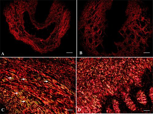
The giant anteater (Myrmecophaga tridactyla) belongs to the superorder Xenarthra and is distributed throughout Central and South America. This animal is listed as ‘vulnerable’ on the International Union for Conservation of Nature's Red List of Threatened Species. Studies on the reproduction of this species are essential because of its peculiarities; however, there is a lack of information on its reproductive aspects and the biotechnologies that can be applied to it. Morphological and histopathological descriptions of the reproductive organs of Myrmecophaga tridactyla are fundamental for determining the general characteristics that could improve our understanding of reproductive disorders in this species. Therefore, this study aimed to perform morphological and histochemical characterisation of the penis of Myrmecophaga tridactyla. For this purpose, we collected six postmortem samples of giant anteater penises. The penis of the giant anteater has no foreskin, two erectile bodies, a paired corpora cavernosa, corpus spongiosum and urethra. Type I (red) and type III (yellow-green) collagen fibres were distributed throughout the stroma and erectile bodies of the penis. Periodic acid-Schiff (PAS)-positive staining was observed in the epithelial cells at the base of the hair follicles, and immunohistochemical immunolabelling for androgen receptors (AR) and oestrogen receptors (ER) was detected in all cells of the transitional epithelium of the penile urethra. These findings suggest that penile urethra cells are sensitive to oestrogen and progesterone.
期刊介绍:
Anatomia, Histologia, Embryologia is a premier international forum for the latest research on descriptive, applied and clinical anatomy, histology, embryology, and related fields. Special emphasis is placed on the links between animal morphology and veterinary and experimental medicine, consequently studies on clinically relevant species will be given priority. The editors welcome papers on medical imaging and anatomical techniques. The journal is of vital interest to clinicians, zoologists, obstetricians, and researchers working in biotechnology. Contributions include reviews, original research articles, short communications and book reviews.
 求助内容:
求助内容: 应助结果提醒方式:


