Sammy Onyancha, Ramin Lonnes, Peter Hollaus, Waldemar Schreiner, Gernot Rohde
{"title":"孤立性肺病变中模拟恶性的分枝杆菌感染:锥束ct引导下的活检病例系列。","authors":"Sammy Onyancha, Ramin Lonnes, Peter Hollaus, Waldemar Schreiner, Gernot Rohde","doi":"10.1002/rcr2.70300","DOIUrl":null,"url":null,"abstract":"<p><p>Solitary pulmonary lesions are often associated with malignancy. Cone beam computed tomography (CBCT) enhances bronchoscopic biopsy accuracy by confirming tool-in-lesion positioning. We present five cases of solitary pulmonary nodules initially suspected to be malignant based on imaging and clinical context. Despite clear tool-in-lesion confirmation via CBCT, initial pathology was non-diagnostic for malignancy. Upon further microbiological analysis, four cases were diagnosed as mycobacterial infections. A fifth case, which underwent surgical resection due to persistent diagnostic uncertainty, was subsequently found to harbour mycobacterial infection; retrospective review of the original biopsy also confirmed this. These cases highlight the importance of including mycobacterial infections such as tuberculoma in the differential diagnosis of solitary pulmonary nodules and stress the need for comprehensive microbiological evaluation in CBCT-confirmed biopsies, especially when histology is non-malignant. Our findings also emphasise the potential diagnostic utility of microbiological tests-including PCR-even prior to histology review when CBCT confirms tool-in-lesion. This approach may prevent unnecessary surgical interventions and associated morbidity.</p>","PeriodicalId":45846,"journal":{"name":"Respirology Case Reports","volume":"13 8","pages":"e70300"},"PeriodicalIF":0.8000,"publicationDate":"2025-08-03","publicationTypes":"Journal Article","fieldsOfStudy":null,"isOpenAccess":false,"openAccessPdf":"https://www.ncbi.nlm.nih.gov/pmc/articles/PMC12318634/pdf/","citationCount":"0","resultStr":"{\"title\":\"Mycobacterial Infections Mimicking Malignancy in Solitary Pulmonary Lesions: A Cone Beam CT-Guided Biopsy Case Series.\",\"authors\":\"Sammy Onyancha, Ramin Lonnes, Peter Hollaus, Waldemar Schreiner, Gernot Rohde\",\"doi\":\"10.1002/rcr2.70300\",\"DOIUrl\":null,\"url\":null,\"abstract\":\"<p><p>Solitary pulmonary lesions are often associated with malignancy. Cone beam computed tomography (CBCT) enhances bronchoscopic biopsy accuracy by confirming tool-in-lesion positioning. We present five cases of solitary pulmonary nodules initially suspected to be malignant based on imaging and clinical context. Despite clear tool-in-lesion confirmation via CBCT, initial pathology was non-diagnostic for malignancy. Upon further microbiological analysis, four cases were diagnosed as mycobacterial infections. A fifth case, which underwent surgical resection due to persistent diagnostic uncertainty, was subsequently found to harbour mycobacterial infection; retrospective review of the original biopsy also confirmed this. These cases highlight the importance of including mycobacterial infections such as tuberculoma in the differential diagnosis of solitary pulmonary nodules and stress the need for comprehensive microbiological evaluation in CBCT-confirmed biopsies, especially when histology is non-malignant. Our findings also emphasise the potential diagnostic utility of microbiological tests-including PCR-even prior to histology review when CBCT confirms tool-in-lesion. This approach may prevent unnecessary surgical interventions and associated morbidity.</p>\",\"PeriodicalId\":45846,\"journal\":{\"name\":\"Respirology Case Reports\",\"volume\":\"13 8\",\"pages\":\"e70300\"},\"PeriodicalIF\":0.8000,\"publicationDate\":\"2025-08-03\",\"publicationTypes\":\"Journal Article\",\"fieldsOfStudy\":null,\"isOpenAccess\":false,\"openAccessPdf\":\"https://www.ncbi.nlm.nih.gov/pmc/articles/PMC12318634/pdf/\",\"citationCount\":\"0\",\"resultStr\":null,\"platform\":\"Semanticscholar\",\"paperid\":null,\"PeriodicalName\":\"Respirology Case Reports\",\"FirstCategoryId\":\"1085\",\"ListUrlMain\":\"https://doi.org/10.1002/rcr2.70300\",\"RegionNum\":0,\"RegionCategory\":null,\"ArticlePicture\":[],\"TitleCN\":null,\"AbstractTextCN\":null,\"PMCID\":null,\"EPubDate\":\"2025/8/1 0:00:00\",\"PubModel\":\"eCollection\",\"JCR\":\"Q4\",\"JCRName\":\"RESPIRATORY SYSTEM\",\"Score\":null,\"Total\":0}","platform":"Semanticscholar","paperid":null,"PeriodicalName":"Respirology Case Reports","FirstCategoryId":"1085","ListUrlMain":"https://doi.org/10.1002/rcr2.70300","RegionNum":0,"RegionCategory":null,"ArticlePicture":[],"TitleCN":null,"AbstractTextCN":null,"PMCID":null,"EPubDate":"2025/8/1 0:00:00","PubModel":"eCollection","JCR":"Q4","JCRName":"RESPIRATORY SYSTEM","Score":null,"Total":0}
Mycobacterial Infections Mimicking Malignancy in Solitary Pulmonary Lesions: A Cone Beam CT-Guided Biopsy Case Series.
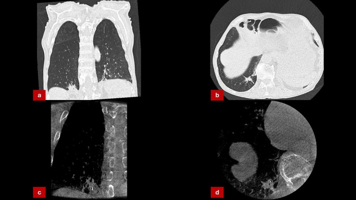
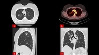
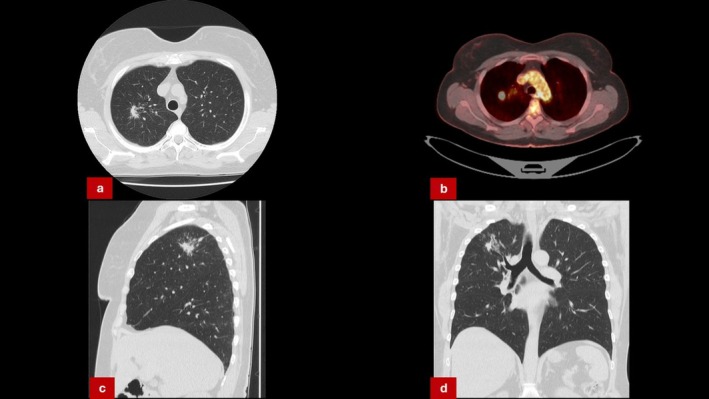
Solitary pulmonary lesions are often associated with malignancy. Cone beam computed tomography (CBCT) enhances bronchoscopic biopsy accuracy by confirming tool-in-lesion positioning. We present five cases of solitary pulmonary nodules initially suspected to be malignant based on imaging and clinical context. Despite clear tool-in-lesion confirmation via CBCT, initial pathology was non-diagnostic for malignancy. Upon further microbiological analysis, four cases were diagnosed as mycobacterial infections. A fifth case, which underwent surgical resection due to persistent diagnostic uncertainty, was subsequently found to harbour mycobacterial infection; retrospective review of the original biopsy also confirmed this. These cases highlight the importance of including mycobacterial infections such as tuberculoma in the differential diagnosis of solitary pulmonary nodules and stress the need for comprehensive microbiological evaluation in CBCT-confirmed biopsies, especially when histology is non-malignant. Our findings also emphasise the potential diagnostic utility of microbiological tests-including PCR-even prior to histology review when CBCT confirms tool-in-lesion. This approach may prevent unnecessary surgical interventions and associated morbidity.
期刊介绍:
Respirology Case Reports is an open-access online journal dedicated to the publication of original clinical case reports, case series, clinical images and clinical videos in all fields of respiratory medicine. The Journal encourages the international exchange between clinicians and researchers of experiences in diagnosing and treating uncommon diseases or diseases with unusual presentations. All manuscripts are peer-reviewed through a streamlined process that aims at providing a rapid turnaround time from submission to publication.

 求助内容:
求助内容: 应助结果提醒方式:


