Yue Cheng, Ran Ren, Yu Xu, Shaofeng Duan, Jilei Zhang, Zhongyuan Bao
{"title":"基于动态增强mri的肿瘤内动力学异质性放射组学模型用于预测乳腺癌分子亚型。","authors":"Yue Cheng, Ran Ren, Yu Xu, Shaofeng Duan, Jilei Zhang, Zhongyuan Bao","doi":"10.3389/fmolb.2025.1635296","DOIUrl":null,"url":null,"abstract":"<p><strong>Objectives: </strong>This study aims to segment intra-tumoral subregions of breast cancer based on kinetic heterogeneity using dynamic contrast-enhanced magnetic resonance imaging (DCE-MRI). It also aims to construct a radiomics model of the whole tumor and washout region to predict molecular subtypes and human epidermal growth factor receptor 2 (HER2) status.</p><p><strong>Methods: </strong>A total of 124 patients with biopsy-confirmed breast cancer were randomly divided into training and test sets in a 7:3 ratio. Quantitative analysis of breast cancer kinetic heterogeneity parameters based on DCE-MRI data was performed, dividing tumors into three subregions (Persistent, Washout, and Plateau) according to the type of voxel-level contrast enhancement. Radiomics features of the washout region and the whole tumor were extracted from the first phase of DCE-MRI enhancement. The area under the receiver operating characteristic curve (AUC) and decision curve analysis (DCA) were used to evaluate the performance of the model.</p><p><strong>Results: </strong>The radiomics model using tumor subregion (washout region) features related to kinetic heterogeneity showed the best performance for differentiating between patients with Luminal, HER2, and HER2 status, with AUC values in the train set of 0.924, 0.876, and 0.816, respectively. Exhibiting an AUC value higher than that obtained with the whole tumor and the kinetic heterogeneity parameters. DCA curves showed that the washout region model was more effective in predicting Luminal and HER2-status subtypes, compared to the whole tumor region model.</p><p><strong>Conclusion: </strong>Radiomics analysis of washout areas from high-resolution DCE-MRI breast scans has the potential to better identify molecular subtypes of breast cancer non-invasively.</p>","PeriodicalId":12465,"journal":{"name":"Frontiers in Molecular Biosciences","volume":"12 ","pages":"1635296"},"PeriodicalIF":3.9000,"publicationDate":"2025-07-18","publicationTypes":"Journal Article","fieldsOfStudy":null,"isOpenAccess":false,"openAccessPdf":"https://www.ncbi.nlm.nih.gov/pmc/articles/PMC12313481/pdf/","citationCount":"0","resultStr":"{\"title\":\"Dynamic contrast-enhanced MRI-based radiomics model of intra-tumoral kinetic heterogeneity for predicting breast cancer molecular subtypes.\",\"authors\":\"Yue Cheng, Ran Ren, Yu Xu, Shaofeng Duan, Jilei Zhang, Zhongyuan Bao\",\"doi\":\"10.3389/fmolb.2025.1635296\",\"DOIUrl\":null,\"url\":null,\"abstract\":\"<p><strong>Objectives: </strong>This study aims to segment intra-tumoral subregions of breast cancer based on kinetic heterogeneity using dynamic contrast-enhanced magnetic resonance imaging (DCE-MRI). It also aims to construct a radiomics model of the whole tumor and washout region to predict molecular subtypes and human epidermal growth factor receptor 2 (HER2) status.</p><p><strong>Methods: </strong>A total of 124 patients with biopsy-confirmed breast cancer were randomly divided into training and test sets in a 7:3 ratio. Quantitative analysis of breast cancer kinetic heterogeneity parameters based on DCE-MRI data was performed, dividing tumors into three subregions (Persistent, Washout, and Plateau) according to the type of voxel-level contrast enhancement. Radiomics features of the washout region and the whole tumor were extracted from the first phase of DCE-MRI enhancement. The area under the receiver operating characteristic curve (AUC) and decision curve analysis (DCA) were used to evaluate the performance of the model.</p><p><strong>Results: </strong>The radiomics model using tumor subregion (washout region) features related to kinetic heterogeneity showed the best performance for differentiating between patients with Luminal, HER2, and HER2 status, with AUC values in the train set of 0.924, 0.876, and 0.816, respectively. Exhibiting an AUC value higher than that obtained with the whole tumor and the kinetic heterogeneity parameters. DCA curves showed that the washout region model was more effective in predicting Luminal and HER2-status subtypes, compared to the whole tumor region model.</p><p><strong>Conclusion: </strong>Radiomics analysis of washout areas from high-resolution DCE-MRI breast scans has the potential to better identify molecular subtypes of breast cancer non-invasively.</p>\",\"PeriodicalId\":12465,\"journal\":{\"name\":\"Frontiers in Molecular Biosciences\",\"volume\":\"12 \",\"pages\":\"1635296\"},\"PeriodicalIF\":3.9000,\"publicationDate\":\"2025-07-18\",\"publicationTypes\":\"Journal Article\",\"fieldsOfStudy\":null,\"isOpenAccess\":false,\"openAccessPdf\":\"https://www.ncbi.nlm.nih.gov/pmc/articles/PMC12313481/pdf/\",\"citationCount\":\"0\",\"resultStr\":null,\"platform\":\"Semanticscholar\",\"paperid\":null,\"PeriodicalName\":\"Frontiers in Molecular Biosciences\",\"FirstCategoryId\":\"99\",\"ListUrlMain\":\"https://doi.org/10.3389/fmolb.2025.1635296\",\"RegionNum\":3,\"RegionCategory\":\"生物学\",\"ArticlePicture\":[],\"TitleCN\":null,\"AbstractTextCN\":null,\"PMCID\":null,\"EPubDate\":\"2025/1/1 0:00:00\",\"PubModel\":\"eCollection\",\"JCR\":\"Q2\",\"JCRName\":\"BIOCHEMISTRY & MOLECULAR BIOLOGY\",\"Score\":null,\"Total\":0}","platform":"Semanticscholar","paperid":null,"PeriodicalName":"Frontiers in Molecular Biosciences","FirstCategoryId":"99","ListUrlMain":"https://doi.org/10.3389/fmolb.2025.1635296","RegionNum":3,"RegionCategory":"生物学","ArticlePicture":[],"TitleCN":null,"AbstractTextCN":null,"PMCID":null,"EPubDate":"2025/1/1 0:00:00","PubModel":"eCollection","JCR":"Q2","JCRName":"BIOCHEMISTRY & MOLECULAR BIOLOGY","Score":null,"Total":0}
Dynamic contrast-enhanced MRI-based radiomics model of intra-tumoral kinetic heterogeneity for predicting breast cancer molecular subtypes.
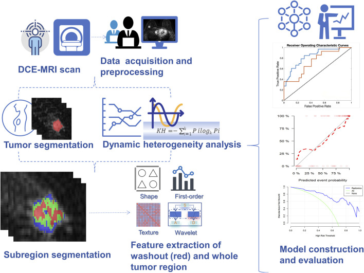
Objectives: This study aims to segment intra-tumoral subregions of breast cancer based on kinetic heterogeneity using dynamic contrast-enhanced magnetic resonance imaging (DCE-MRI). It also aims to construct a radiomics model of the whole tumor and washout region to predict molecular subtypes and human epidermal growth factor receptor 2 (HER2) status.
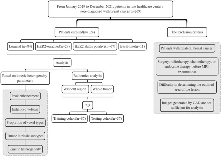
Methods: A total of 124 patients with biopsy-confirmed breast cancer were randomly divided into training and test sets in a 7:3 ratio. Quantitative analysis of breast cancer kinetic heterogeneity parameters based on DCE-MRI data was performed, dividing tumors into three subregions (Persistent, Washout, and Plateau) according to the type of voxel-level contrast enhancement. Radiomics features of the washout region and the whole tumor were extracted from the first phase of DCE-MRI enhancement. The area under the receiver operating characteristic curve (AUC) and decision curve analysis (DCA) were used to evaluate the performance of the model.
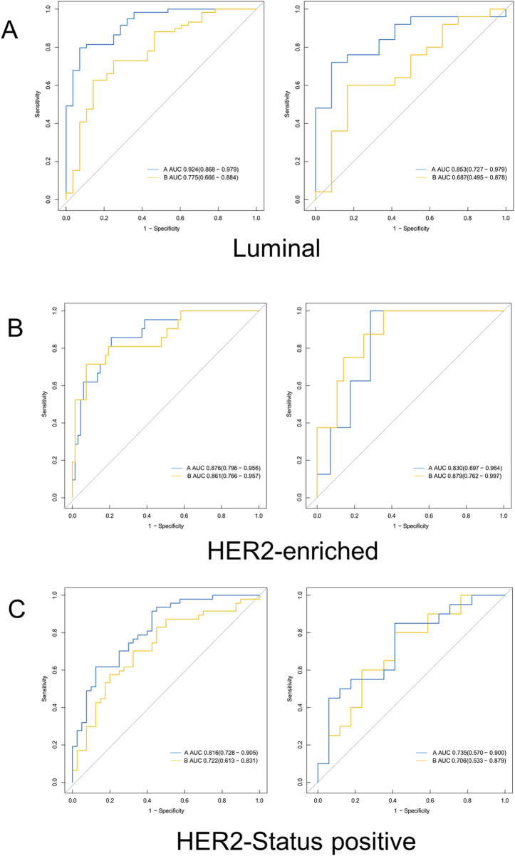
Results: The radiomics model using tumor subregion (washout region) features related to kinetic heterogeneity showed the best performance for differentiating between patients with Luminal, HER2, and HER2 status, with AUC values in the train set of 0.924, 0.876, and 0.816, respectively. Exhibiting an AUC value higher than that obtained with the whole tumor and the kinetic heterogeneity parameters. DCA curves showed that the washout region model was more effective in predicting Luminal and HER2-status subtypes, compared to the whole tumor region model.
Conclusion: Radiomics analysis of washout areas from high-resolution DCE-MRI breast scans has the potential to better identify molecular subtypes of breast cancer non-invasively.
期刊介绍:
Much of contemporary investigation in the life sciences is devoted to the molecular-scale understanding of the relationships between genes and the environment — in particular, dynamic alterations in the levels, modifications, and interactions of cellular effectors, including proteins. Frontiers in Molecular Biosciences offers an international publication platform for basic as well as applied research; we encourage contributions spanning both established and emerging areas of biology. To this end, the journal draws from empirical disciplines such as structural biology, enzymology, biochemistry, and biophysics, capitalizing as well on the technological advancements that have enabled metabolomics and proteomics measurements in massively parallel throughput, and the development of robust and innovative computational biology strategies. We also recognize influences from medicine and technology, welcoming studies in molecular genetics, molecular diagnostics and therapeutics, and nanotechnology.
Our ultimate objective is the comprehensive illustration of the molecular mechanisms regulating proteins, nucleic acids, carbohydrates, lipids, and small metabolites in organisms across all branches of life.
In addition to interesting new findings, techniques, and applications, Frontiers in Molecular Biosciences will consider new testable hypotheses to inspire different perspectives and stimulate scientific dialogue. The integration of in silico, in vitro, and in vivo approaches will benefit endeavors across all domains of the life sciences.
 求助内容:
求助内容: 应助结果提醒方式:


