{"title":"使用IVUS和OFDI进行耳蜗内成像:尸体的可行性研究","authors":"Ayu Akazawa, Takeshi Fujita, Jun Yokoi, Natsumi Uehara, Mie Kubo, Akinobu Kakigi, Ken-ichi Nibu","doi":"10.1002/lio2.70224","DOIUrl":null,"url":null,"abstract":"<div>\n \n \n <section>\n \n <h3> Objective</h3>\n \n <p>This study evaluates the feasibility of optical frequency domain imaging (OFDI) and intravascular ultrasound (IVUS) for intracochlear imaging in human cadaveric specimens. We compare their resolution, depth penetration, and ability to identify key cochlear structures and pathological conditions.</p>\n </section>\n \n <section>\n \n <h3> Methods</h3>\n \n <p>Human cadaveric temporal bones were prepared, and OFDI and IVUS probes were inserted through the round window into the scala tympani. OFDI imaging was performed using the LUNAWAVE system (Terumo, Tokyo, Japan), while IVUS imaging was conducted using the VISICUBE system (Terumo, Tokyo, Japan) with 60 and 40 MHz probes. The probe tips were trimmed to optimize insertion and imaging. Imaging results were compared with histological sections.</p>\n </section>\n \n <section>\n \n <h3> Results</h3>\n \n <p>OFDI provided high-resolution visualization of the cochlear basal turn, allowing clear identification of the basilar membrane, Reissner's membrane, scala media, and the organ of Corti. IVUS exhibited lower resolution than OFDI but allowed for deeper tissue penetration. The 60 MHz IVUS probe provided higher resolution than the 40 MHz probe, while the 40 MHz probe offered a wider field of view due to greater penetration depth and lower attenuation. Both OFDI and IVUS successfully detected basilar membrane disruptions, a known complication in cochlear implant surgery.</p>\n </section>\n \n <section>\n \n <h3> Conclusion</h3>\n \n <p>OFDI and IVUS demonstrated feasibility for intracochlear imaging, with OFDI offering superior resolution and IVUS providing greater penetration. This is the first study to apply IVUS for intracochlear imaging, supporting its potential role in intraoperative monitoring and cochlear pathology assessment.</p>\n </section>\n \n <section>\n \n <h3> Level of Evidence</h3>\n \n <p>N/A.</p>\n </section>\n </div>","PeriodicalId":48529,"journal":{"name":"Laryngoscope Investigative Otolaryngology","volume":"10 4","pages":""},"PeriodicalIF":1.7000,"publicationDate":"2025-08-05","publicationTypes":"Journal Article","fieldsOfStudy":null,"isOpenAccess":false,"openAccessPdf":"https://onlinelibrary.wiley.com/doi/epdf/10.1002/lio2.70224","citationCount":"0","resultStr":"{\"title\":\"Intracochlear Imaging Using IVUS and OFDI: A Cadaveric Feasibility Study\",\"authors\":\"Ayu Akazawa, Takeshi Fujita, Jun Yokoi, Natsumi Uehara, Mie Kubo, Akinobu Kakigi, Ken-ichi Nibu\",\"doi\":\"10.1002/lio2.70224\",\"DOIUrl\":null,\"url\":null,\"abstract\":\"<div>\\n \\n \\n <section>\\n \\n <h3> Objective</h3>\\n \\n <p>This study evaluates the feasibility of optical frequency domain imaging (OFDI) and intravascular ultrasound (IVUS) for intracochlear imaging in human cadaveric specimens. We compare their resolution, depth penetration, and ability to identify key cochlear structures and pathological conditions.</p>\\n </section>\\n \\n <section>\\n \\n <h3> Methods</h3>\\n \\n <p>Human cadaveric temporal bones were prepared, and OFDI and IVUS probes were inserted through the round window into the scala tympani. OFDI imaging was performed using the LUNAWAVE system (Terumo, Tokyo, Japan), while IVUS imaging was conducted using the VISICUBE system (Terumo, Tokyo, Japan) with 60 and 40 MHz probes. The probe tips were trimmed to optimize insertion and imaging. Imaging results were compared with histological sections.</p>\\n </section>\\n \\n <section>\\n \\n <h3> Results</h3>\\n \\n <p>OFDI provided high-resolution visualization of the cochlear basal turn, allowing clear identification of the basilar membrane, Reissner's membrane, scala media, and the organ of Corti. IVUS exhibited lower resolution than OFDI but allowed for deeper tissue penetration. The 60 MHz IVUS probe provided higher resolution than the 40 MHz probe, while the 40 MHz probe offered a wider field of view due to greater penetration depth and lower attenuation. Both OFDI and IVUS successfully detected basilar membrane disruptions, a known complication in cochlear implant surgery.</p>\\n </section>\\n \\n <section>\\n \\n <h3> Conclusion</h3>\\n \\n <p>OFDI and IVUS demonstrated feasibility for intracochlear imaging, with OFDI offering superior resolution and IVUS providing greater penetration. This is the first study to apply IVUS for intracochlear imaging, supporting its potential role in intraoperative monitoring and cochlear pathology assessment.</p>\\n </section>\\n \\n <section>\\n \\n <h3> Level of Evidence</h3>\\n \\n <p>N/A.</p>\\n </section>\\n </div>\",\"PeriodicalId\":48529,\"journal\":{\"name\":\"Laryngoscope Investigative Otolaryngology\",\"volume\":\"10 4\",\"pages\":\"\"},\"PeriodicalIF\":1.7000,\"publicationDate\":\"2025-08-05\",\"publicationTypes\":\"Journal Article\",\"fieldsOfStudy\":null,\"isOpenAccess\":false,\"openAccessPdf\":\"https://onlinelibrary.wiley.com/doi/epdf/10.1002/lio2.70224\",\"citationCount\":\"0\",\"resultStr\":null,\"platform\":\"Semanticscholar\",\"paperid\":null,\"PeriodicalName\":\"Laryngoscope Investigative Otolaryngology\",\"FirstCategoryId\":\"3\",\"ListUrlMain\":\"https://onlinelibrary.wiley.com/doi/10.1002/lio2.70224\",\"RegionNum\":4,\"RegionCategory\":\"医学\",\"ArticlePicture\":[],\"TitleCN\":null,\"AbstractTextCN\":null,\"PMCID\":null,\"EPubDate\":\"\",\"PubModel\":\"\",\"JCR\":\"Q2\",\"JCRName\":\"OTORHINOLARYNGOLOGY\",\"Score\":null,\"Total\":0}","platform":"Semanticscholar","paperid":null,"PeriodicalName":"Laryngoscope Investigative Otolaryngology","FirstCategoryId":"3","ListUrlMain":"https://onlinelibrary.wiley.com/doi/10.1002/lio2.70224","RegionNum":4,"RegionCategory":"医学","ArticlePicture":[],"TitleCN":null,"AbstractTextCN":null,"PMCID":null,"EPubDate":"","PubModel":"","JCR":"Q2","JCRName":"OTORHINOLARYNGOLOGY","Score":null,"Total":0}
引用次数: 0
摘要
目的探讨光学频域成像(OFDI)和血管内超声(IVUS)在人尸体耳蜗内成像中的可行性。我们比较了它们的分辨率,深度穿透,以及识别关键耳蜗结构和病理状况的能力。方法制备人尸体颞骨,经圆窗置入OFDI和IVUS探针。OFDI成像使用LUNAWAVE系统(Terumo, Tokyo, Japan), IVUS成像使用VISICUBE系统(Terumo, Tokyo, Japan), 60和40 MHz探头。修剪探针尖端以优化插入和成像。影像学结果与组织学切片比较。结果OFDI提供了高分辨率的耳蜗基底转的可视化,可以清楚地识别基底膜、Reissner膜、中膜和Corti器官。IVUS的分辨率比OFDI低,但可以穿透更深的组织。60 MHz IVUS探头比40 MHz探头提供更高的分辨率,而40 MHz探头由于更大的穿透深度和更低的衰减而提供更宽的视野。OFDI和IVUS都成功地检测到基底膜破坏,这是人工耳蜗手术中已知的并发症。结论OFDI和IVUS在耳蜗内成像具有可行性,OFDI分辨率高,IVUS穿透力强。这是首次将IVUS应用于耳蜗内成像的研究,支持其在术中监测和耳蜗病理评估中的潜在作用。证据水平:无。
Intracochlear Imaging Using IVUS and OFDI: A Cadaveric Feasibility Study
Objective
This study evaluates the feasibility of optical frequency domain imaging (OFDI) and intravascular ultrasound (IVUS) for intracochlear imaging in human cadaveric specimens. We compare their resolution, depth penetration, and ability to identify key cochlear structures and pathological conditions.
Methods
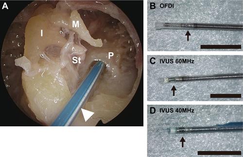
Human cadaveric temporal bones were prepared, and OFDI and IVUS probes were inserted through the round window into the scala tympani. OFDI imaging was performed using the LUNAWAVE system (Terumo, Tokyo, Japan), while IVUS imaging was conducted using the VISICUBE system (Terumo, Tokyo, Japan) with 60 and 40 MHz probes. The probe tips were trimmed to optimize insertion and imaging. Imaging results were compared with histological sections.
Results
OFDI provided high-resolution visualization of the cochlear basal turn, allowing clear identification of the basilar membrane, Reissner's membrane, scala media, and the organ of Corti. IVUS exhibited lower resolution than OFDI but allowed for deeper tissue penetration. The 60 MHz IVUS probe provided higher resolution than the 40 MHz probe, while the 40 MHz probe offered a wider field of view due to greater penetration depth and lower attenuation. Both OFDI and IVUS successfully detected basilar membrane disruptions, a known complication in cochlear implant surgery.
Conclusion
OFDI and IVUS demonstrated feasibility for intracochlear imaging, with OFDI offering superior resolution and IVUS providing greater penetration. This is the first study to apply IVUS for intracochlear imaging, supporting its potential role in intraoperative monitoring and cochlear pathology assessment.
 求助内容:
求助内容: 应助结果提醒方式:


