{"title":"3d打印聚乳酸作为骨替代品的评估:在大鼠模型中的动物研究","authors":"Velayudhan Ashok, Mohanraj Karthik Ganesh, Subhabrata Maiti, Deepak Nallaswamy, Artak Heboyan","doi":"10.1002/cre2.70201","DOIUrl":null,"url":null,"abstract":"<div>\n \n \n <section>\n \n <h3> Objectives</h3>\n \n <p>Bone repair and regeneration are important processes for treating bone defects and injuries. However, traditional bone grafts like autografts and allografts have limitations, such as complications at the donor site and immune rejection. As a result, there is growing interest in using polylactic acid (PLA), a biodegradable and biocompatible material, as a synthetic bone substitute. This study aims to evaluate the effectiveness of 3D-printed PLA scaffolds as bone substitutes using a rat model.</p>\n </section>\n \n <section>\n \n <h3> Material and Methods</h3>\n \n <p>PLA scaffolds with dimensions of 2 × 2 × 4 mm and 2 × 2 × 8 mm were fabricated using the CUBEX-TRIO 3D printer. Twelve male Wistar rats were divided into four groups based on defect size (4 and 8 mm) and observation period (4 weeks and 8 weeks). The surgical procedures involved creating discontinuity defects in the rats' zygoma and implanting PLA scaffolds that were stabilized with a bio-membrane. Bone regeneration was assessed through radiographic analysis and histological examination.</p>\n </section>\n \n <section>\n \n <h3> Results</h3>\n \n <p>Radiographic analysis confirmed the formation of bone in the grafted regions. Histological analysis revealed connective tissue formation at the defect edges and scaffold surface at both 4 and 8 weeks. In the 4 mm defect group, the transformation of connective tissue into chondrocytes and endochondral ossification was observed at 8 weeks, indicating successful bone regeneration. However, in the 8 mm defect group, bone formation was not as evident, suggesting limitations in the osteoinductive potential of PLA scaffolds for larger defects.</p>\n </section>\n \n <section>\n \n <h3> Conclusions</h3>\n \n <p>The 3D-printed PLA scaffolds show promise as bone substitutes for small to moderate-sized defects due to their effective biocompatibility and osteoinductive potential. Further studies are needed to optimize their performance for larger defects, potentially enhancing their clinical application in bone repair and regeneration.</p>\n </section>\n </div>","PeriodicalId":10203,"journal":{"name":"Clinical and Experimental Dental Research","volume":"11 4","pages":""},"PeriodicalIF":2.2000,"publicationDate":"2025-08-05","publicationTypes":"Journal Article","fieldsOfStudy":null,"isOpenAccess":false,"openAccessPdf":"https://onlinelibrary.wiley.com/doi/epdf/10.1002/cre2.70201","citationCount":"0","resultStr":"{\"title\":\"Evaluation of 3D-Printed Polylactic Acid as a Bone Substitute: An Animal Study in a Rat Model\",\"authors\":\"Velayudhan Ashok, Mohanraj Karthik Ganesh, Subhabrata Maiti, Deepak Nallaswamy, Artak Heboyan\",\"doi\":\"10.1002/cre2.70201\",\"DOIUrl\":null,\"url\":null,\"abstract\":\"<div>\\n \\n \\n <section>\\n \\n <h3> Objectives</h3>\\n \\n <p>Bone repair and regeneration are important processes for treating bone defects and injuries. However, traditional bone grafts like autografts and allografts have limitations, such as complications at the donor site and immune rejection. As a result, there is growing interest in using polylactic acid (PLA), a biodegradable and biocompatible material, as a synthetic bone substitute. This study aims to evaluate the effectiveness of 3D-printed PLA scaffolds as bone substitutes using a rat model.</p>\\n </section>\\n \\n <section>\\n \\n <h3> Material and Methods</h3>\\n \\n <p>PLA scaffolds with dimensions of 2 × 2 × 4 mm and 2 × 2 × 8 mm were fabricated using the CUBEX-TRIO 3D printer. Twelve male Wistar rats were divided into four groups based on defect size (4 and 8 mm) and observation period (4 weeks and 8 weeks). The surgical procedures involved creating discontinuity defects in the rats' zygoma and implanting PLA scaffolds that were stabilized with a bio-membrane. Bone regeneration was assessed through radiographic analysis and histological examination.</p>\\n </section>\\n \\n <section>\\n \\n <h3> Results</h3>\\n \\n <p>Radiographic analysis confirmed the formation of bone in the grafted regions. Histological analysis revealed connective tissue formation at the defect edges and scaffold surface at both 4 and 8 weeks. In the 4 mm defect group, the transformation of connective tissue into chondrocytes and endochondral ossification was observed at 8 weeks, indicating successful bone regeneration. However, in the 8 mm defect group, bone formation was not as evident, suggesting limitations in the osteoinductive potential of PLA scaffolds for larger defects.</p>\\n </section>\\n \\n <section>\\n \\n <h3> Conclusions</h3>\\n \\n <p>The 3D-printed PLA scaffolds show promise as bone substitutes for small to moderate-sized defects due to their effective biocompatibility and osteoinductive potential. Further studies are needed to optimize their performance for larger defects, potentially enhancing their clinical application in bone repair and regeneration.</p>\\n </section>\\n </div>\",\"PeriodicalId\":10203,\"journal\":{\"name\":\"Clinical and Experimental Dental Research\",\"volume\":\"11 4\",\"pages\":\"\"},\"PeriodicalIF\":2.2000,\"publicationDate\":\"2025-08-05\",\"publicationTypes\":\"Journal Article\",\"fieldsOfStudy\":null,\"isOpenAccess\":false,\"openAccessPdf\":\"https://onlinelibrary.wiley.com/doi/epdf/10.1002/cre2.70201\",\"citationCount\":\"0\",\"resultStr\":null,\"platform\":\"Semanticscholar\",\"paperid\":null,\"PeriodicalName\":\"Clinical and Experimental Dental Research\",\"FirstCategoryId\":\"1085\",\"ListUrlMain\":\"https://onlinelibrary.wiley.com/doi/10.1002/cre2.70201\",\"RegionNum\":0,\"RegionCategory\":null,\"ArticlePicture\":[],\"TitleCN\":null,\"AbstractTextCN\":null,\"PMCID\":null,\"EPubDate\":\"\",\"PubModel\":\"\",\"JCR\":\"Q3\",\"JCRName\":\"DENTISTRY, ORAL SURGERY & MEDICINE\",\"Score\":null,\"Total\":0}","platform":"Semanticscholar","paperid":null,"PeriodicalName":"Clinical and Experimental Dental Research","FirstCategoryId":"1085","ListUrlMain":"https://onlinelibrary.wiley.com/doi/10.1002/cre2.70201","RegionNum":0,"RegionCategory":null,"ArticlePicture":[],"TitleCN":null,"AbstractTextCN":null,"PMCID":null,"EPubDate":"","PubModel":"","JCR":"Q3","JCRName":"DENTISTRY, ORAL SURGERY & MEDICINE","Score":null,"Total":0}
Evaluation of 3D-Printed Polylactic Acid as a Bone Substitute: An Animal Study in a Rat Model
Objectives
Bone repair and regeneration are important processes for treating bone defects and injuries. However, traditional bone grafts like autografts and allografts have limitations, such as complications at the donor site and immune rejection. As a result, there is growing interest in using polylactic acid (PLA), a biodegradable and biocompatible material, as a synthetic bone substitute. This study aims to evaluate the effectiveness of 3D-printed PLA scaffolds as bone substitutes using a rat model.
Material and Methods
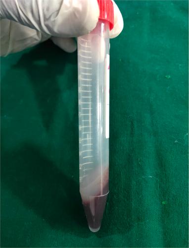
PLA scaffolds with dimensions of 2 × 2 × 4 mm and 2 × 2 × 8 mm were fabricated using the CUBEX-TRIO 3D printer. Twelve male Wistar rats were divided into four groups based on defect size (4 and 8 mm) and observation period (4 weeks and 8 weeks). The surgical procedures involved creating discontinuity defects in the rats' zygoma and implanting PLA scaffolds that were stabilized with a bio-membrane. Bone regeneration was assessed through radiographic analysis and histological examination.
Results
Radiographic analysis confirmed the formation of bone in the grafted regions. Histological analysis revealed connective tissue formation at the defect edges and scaffold surface at both 4 and 8 weeks. In the 4 mm defect group, the transformation of connective tissue into chondrocytes and endochondral ossification was observed at 8 weeks, indicating successful bone regeneration. However, in the 8 mm defect group, bone formation was not as evident, suggesting limitations in the osteoinductive potential of PLA scaffolds for larger defects.
Conclusions
The 3D-printed PLA scaffolds show promise as bone substitutes for small to moderate-sized defects due to their effective biocompatibility and osteoinductive potential. Further studies are needed to optimize their performance for larger defects, potentially enhancing their clinical application in bone repair and regeneration.
期刊介绍:
Clinical and Experimental Dental Research aims to provide open access peer-reviewed publications of high scientific quality representing original clinical, diagnostic or experimental work within all disciplines and fields of oral medicine and dentistry. The scope of Clinical and Experimental Dental Research comprises original research material on the anatomy, physiology and pathology of oro-facial, oro-pharyngeal and maxillofacial tissues, and functions and dysfunctions within the stomatognathic system, and the epidemiology, aetiology, prevention, diagnosis, prognosis and therapy of diseases and conditions that have an effect on the homeostasis of the mouth, jaws, and closely associated structures, as well as the healing and regeneration and the clinical aspects of replacement of hard and soft tissues with biomaterials, and the rehabilitation of stomatognathic functions. Studies that bring new knowledge on how to advance health on the individual or public health levels, including interactions between oral and general health and ill-health are welcome.

 求助内容:
求助内容: 应助结果提醒方式:


