Jakob Pansell, Tina Thomsson, Emelie Eng, Andreas Wallin, Mohammad Hirzallah
{"title":"视神经鞘直径:应测量哪个轴?","authors":"Jakob Pansell, Tina Thomsson, Emelie Eng, Andreas Wallin, Mohammad Hirzallah","doi":"10.1111/jon.70076","DOIUrl":null,"url":null,"abstract":"<div>\n \n \n <section>\n \n <h3> Background and purpose</h3>\n \n <p>Optic nerve sheath diameter (ONSD) measured by point-of-care ultrasound (POCUS) is a commonly used non-invasive estimate of intracranial pressure (ICP). However, methodological variations limit standardization of this promising technique. Different imaging axes are identified as one such source of ONSD POCUS methodological variation. This study aimed to evaluate the agreement and diagnostic accuracy for elevated ICP between transverse and sagittal ONSD measurements.</p>\n </section>\n \n <section>\n \n <h3> Methods</h3>\n \n <p>We conducted a post-hoc analysis of 213 intensive care patients from three prior studies. ONSD was measured in both transverse and sagittal planes, externally (ONSDext) and internally (ONSDint) of the dura mater. Agreement was assessed using Lin's concordance correlation coefficient (CCC). Diagnostic accuracy for detecting ICP ≥20 mmHg was evaluated using area under the receiver operator characteristics curve (AUROC) analyses in a subset of 139 patients.</p>\n </section>\n \n <section>\n \n <h3> Results</h3>\n \n <p>Sagittal ONSD was significantly larger than transverse ONSD (median difference 0.2 mm, p<0.001). Agreement between transverse and sagittal ONSD was poor (CCC: 0.65 and 0.70 for right and left side ONSDext, and 0.43 and 0.51 for right and left side ONSDint). No significant differences in AUROC were found between transverse, sagittal, or averaged measurements.</p>\n </section>\n \n <section>\n \n <h3> Conclusions</h3>\n \n <p>Transverse and sagittal ONSD measurements are not interchangeable due to significant differences and poor agreement. Diagnostic accuracy was similar across methods. These findings support continued use of transverse measurement as the standard approach for ONSD POCUS in clinical practice.</p>\n </section>\n </div>","PeriodicalId":16399,"journal":{"name":"Journal of Neuroimaging","volume":"35 4","pages":""},"PeriodicalIF":2.3000,"publicationDate":"2025-08-03","publicationTypes":"Journal Article","fieldsOfStudy":null,"isOpenAccess":false,"openAccessPdf":"https://onlinelibrary.wiley.com/doi/epdf/10.1111/jon.70076","citationCount":"0","resultStr":"{\"title\":\"Optic Nerve Sheath Diameter: Which Axis Should Be Measured?\",\"authors\":\"Jakob Pansell, Tina Thomsson, Emelie Eng, Andreas Wallin, Mohammad Hirzallah\",\"doi\":\"10.1111/jon.70076\",\"DOIUrl\":null,\"url\":null,\"abstract\":\"<div>\\n \\n \\n <section>\\n \\n <h3> Background and purpose</h3>\\n \\n <p>Optic nerve sheath diameter (ONSD) measured by point-of-care ultrasound (POCUS) is a commonly used non-invasive estimate of intracranial pressure (ICP). However, methodological variations limit standardization of this promising technique. Different imaging axes are identified as one such source of ONSD POCUS methodological variation. This study aimed to evaluate the agreement and diagnostic accuracy for elevated ICP between transverse and sagittal ONSD measurements.</p>\\n </section>\\n \\n <section>\\n \\n <h3> Methods</h3>\\n \\n <p>We conducted a post-hoc analysis of 213 intensive care patients from three prior studies. ONSD was measured in both transverse and sagittal planes, externally (ONSDext) and internally (ONSDint) of the dura mater. Agreement was assessed using Lin's concordance correlation coefficient (CCC). Diagnostic accuracy for detecting ICP ≥20 mmHg was evaluated using area under the receiver operator characteristics curve (AUROC) analyses in a subset of 139 patients.</p>\\n </section>\\n \\n <section>\\n \\n <h3> Results</h3>\\n \\n <p>Sagittal ONSD was significantly larger than transverse ONSD (median difference 0.2 mm, p<0.001). Agreement between transverse and sagittal ONSD was poor (CCC: 0.65 and 0.70 for right and left side ONSDext, and 0.43 and 0.51 for right and left side ONSDint). No significant differences in AUROC were found between transverse, sagittal, or averaged measurements.</p>\\n </section>\\n \\n <section>\\n \\n <h3> Conclusions</h3>\\n \\n <p>Transverse and sagittal ONSD measurements are not interchangeable due to significant differences and poor agreement. Diagnostic accuracy was similar across methods. These findings support continued use of transverse measurement as the standard approach for ONSD POCUS in clinical practice.</p>\\n </section>\\n </div>\",\"PeriodicalId\":16399,\"journal\":{\"name\":\"Journal of Neuroimaging\",\"volume\":\"35 4\",\"pages\":\"\"},\"PeriodicalIF\":2.3000,\"publicationDate\":\"2025-08-03\",\"publicationTypes\":\"Journal Article\",\"fieldsOfStudy\":null,\"isOpenAccess\":false,\"openAccessPdf\":\"https://onlinelibrary.wiley.com/doi/epdf/10.1111/jon.70076\",\"citationCount\":\"0\",\"resultStr\":null,\"platform\":\"Semanticscholar\",\"paperid\":null,\"PeriodicalName\":\"Journal of Neuroimaging\",\"FirstCategoryId\":\"3\",\"ListUrlMain\":\"https://onlinelibrary.wiley.com/doi/10.1111/jon.70076\",\"RegionNum\":4,\"RegionCategory\":\"医学\",\"ArticlePicture\":[],\"TitleCN\":null,\"AbstractTextCN\":null,\"PMCID\":null,\"EPubDate\":\"\",\"PubModel\":\"\",\"JCR\":\"Q3\",\"JCRName\":\"CLINICAL NEUROLOGY\",\"Score\":null,\"Total\":0}","platform":"Semanticscholar","paperid":null,"PeriodicalName":"Journal of Neuroimaging","FirstCategoryId":"3","ListUrlMain":"https://onlinelibrary.wiley.com/doi/10.1111/jon.70076","RegionNum":4,"RegionCategory":"医学","ArticlePicture":[],"TitleCN":null,"AbstractTextCN":null,"PMCID":null,"EPubDate":"","PubModel":"","JCR":"Q3","JCRName":"CLINICAL NEUROLOGY","Score":null,"Total":0}
引用次数: 0
摘要
背景与目的视神经鞘直径(ONSD)是一种常用的无创颅内压(ICP)测量方法。然而,方法上的差异限制了这项有前途的技术的标准化。不同的成像轴被认为是ONSD POCUS方法差异的一个来源。本研究旨在评估横向和矢状面ONSD测量之间ICP升高的一致性和诊断准确性。方法:我们对之前三项研究中的213例重症监护患者进行了事后分析。在横切面和矢状面,硬脑膜外部(ONSDext)和内部(ONSDint)测量ONSD。采用Lin’s一致性相关系数(CCC)评价一致性。对139例患者进行受试者操作特征曲线下面积(AUROC)分析,评估检测ICP≥20 mmHg的诊断准确性。结果矢状面ONSD明显大于横面ONSD(中位数差0.2 mm, p<0.001)。横向和矢状面ONSD的一致性较差(左右两侧ONSDext的CCC分别为0.65和0.70,左右两侧ONSDint的CCC分别为0.43和0.51)。横切面、矢状面或平均测量的AUROC无显著差异。结论横切面和矢状面ONSD测量结果差异显著且一致性差,不能互换。不同方法的诊断准确性相似。这些发现支持在临床实践中继续使用横向测量作为ONSD POCUS的标准方法。
Optic Nerve Sheath Diameter: Which Axis Should Be Measured?
Background and purpose
Optic nerve sheath diameter (ONSD) measured by point-of-care ultrasound (POCUS) is a commonly used non-invasive estimate of intracranial pressure (ICP). However, methodological variations limit standardization of this promising technique. Different imaging axes are identified as one such source of ONSD POCUS methodological variation. This study aimed to evaluate the agreement and diagnostic accuracy for elevated ICP between transverse and sagittal ONSD measurements.
Methods
We conducted a post-hoc analysis of 213 intensive care patients from three prior studies. ONSD was measured in both transverse and sagittal planes, externally (ONSDext) and internally (ONSDint) of the dura mater. Agreement was assessed using Lin's concordance correlation coefficient (CCC). Diagnostic accuracy for detecting ICP ≥20 mmHg was evaluated using area under the receiver operator characteristics curve (AUROC) analyses in a subset of 139 patients.
Results
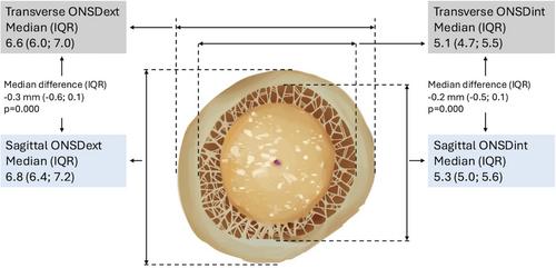
Sagittal ONSD was significantly larger than transverse ONSD (median difference 0.2 mm, p<0.001). Agreement between transverse and sagittal ONSD was poor (CCC: 0.65 and 0.70 for right and left side ONSDext, and 0.43 and 0.51 for right and left side ONSDint). No significant differences in AUROC were found between transverse, sagittal, or averaged measurements.
Conclusions
Transverse and sagittal ONSD measurements are not interchangeable due to significant differences and poor agreement. Diagnostic accuracy was similar across methods. These findings support continued use of transverse measurement as the standard approach for ONSD POCUS in clinical practice.
期刊介绍:
Start reading the Journal of Neuroimaging to learn the latest neurological imaging techniques. The peer-reviewed research is written in a practical clinical context, giving you the information you need on:
MRI
CT
Carotid Ultrasound and TCD
SPECT
PET
Endovascular Surgical Neuroradiology
Functional MRI
Xenon CT
and other new and upcoming neuroscientific modalities.The Journal of Neuroimaging addresses the full spectrum of human nervous system disease, including stroke, neoplasia, degenerating and demyelinating disease, epilepsy, tumors, lesions, infectious disease, cerebral vascular arterial diseases, toxic-metabolic disease, psychoses, dementias, heredo-familial disease, and trauma.Offering original research, review articles, case reports, neuroimaging CPCs, and evaluations of instruments and technology relevant to the nervous system, the Journal of Neuroimaging focuses on useful clinical developments and applications, tested techniques and interpretations, patient care, diagnostics, and therapeutics. Start reading today!

 求助内容:
求助内容: 应助结果提醒方式:


