{"title":"应用扫描源光学相干断层扫描血管造影评估早期糖尿病视网膜病变的视网膜和脉络膜结构及微血管改变。","authors":"Jin Jiang, Xiaole Wang, Hongjun Bian","doi":"10.1371/journal.pone.0327770","DOIUrl":null,"url":null,"abstract":"<p><strong>Objective: </strong>It was to assess changes in structural parameters in early diabetic retinopathy (DR).</p><p><strong>Materials and methodologies: </strong>This study is a retrospective analysis that included patients with early DR admitted to the Affiliated Third Hospital of Nantong University from January 2024 to December 2024. The participants were divided into the non-DR group (NDR group) and the non-proliferative DR group (NPDR group, which included mild, moderate, and severe subgroups) using swept-source optical coherence tomography angiography (SS-OCTA) technology. One-way analysis of variance (ANOVA) and the Kruskal-Wallis test were used to compare parameter differences among the groups.</p><p><strong>Results: </strong>A total of 208 diabetic patients were included (55 in the NDR group, 153 in the NPDR group) and 51 healthy controls. The results showed that the FAZ area in the NPDR group was significantly larger than that in the control group (CG) (mean difference: +0.38 ± 0.10 mm2, 95% CI [0.25-0.51], P < 0.001), and it was positively correlated with disease severity (trend test P < 0.001). Relative to the CG, NDR group and various stages of NPDR group exhibited greatly lower values in choroidal vascular index (CVI), peripapillary vascular density (ppVD), peripapillary retinal nerve fiber layer thickness (pRNFL), vascular density (VD) in both the superficial and deep retinal vascular complexes, total perfusion area (PA), small vessel density (SVD), disc area, vascular density (FD300) within a 300 µm radius of the foveal center, and capillary plexus blood flow density (P<0.05). NPDR group showed progressively lower values than NDR group, with severity increasing as the condition worsened (P<0.05).</p><p><strong>Conclusion: </strong>SS-OCTA can effectively monitor changes in structural parameters and serves as a valuable tool for evaluating the progression of early DR.</p>","PeriodicalId":20189,"journal":{"name":"PLoS ONE","volume":"20 8","pages":"e0327770"},"PeriodicalIF":2.6000,"publicationDate":"2025-08-01","publicationTypes":"Journal Article","fieldsOfStudy":null,"isOpenAccess":false,"openAccessPdf":"https://www.ncbi.nlm.nih.gov/pmc/articles/PMC12316237/pdf/","citationCount":"0","resultStr":"{\"title\":\"Assessment of retinal and choroidal structural and microvascular changes in early diabetic retinopathy using swept-source optical coherence tomography angiography.\",\"authors\":\"Jin Jiang, Xiaole Wang, Hongjun Bian\",\"doi\":\"10.1371/journal.pone.0327770\",\"DOIUrl\":null,\"url\":null,\"abstract\":\"<p><strong>Objective: </strong>It was to assess changes in structural parameters in early diabetic retinopathy (DR).</p><p><strong>Materials and methodologies: </strong>This study is a retrospective analysis that included patients with early DR admitted to the Affiliated Third Hospital of Nantong University from January 2024 to December 2024. The participants were divided into the non-DR group (NDR group) and the non-proliferative DR group (NPDR group, which included mild, moderate, and severe subgroups) using swept-source optical coherence tomography angiography (SS-OCTA) technology. One-way analysis of variance (ANOVA) and the Kruskal-Wallis test were used to compare parameter differences among the groups.</p><p><strong>Results: </strong>A total of 208 diabetic patients were included (55 in the NDR group, 153 in the NPDR group) and 51 healthy controls. The results showed that the FAZ area in the NPDR group was significantly larger than that in the control group (CG) (mean difference: +0.38 ± 0.10 mm2, 95% CI [0.25-0.51], P < 0.001), and it was positively correlated with disease severity (trend test P < 0.001). Relative to the CG, NDR group and various stages of NPDR group exhibited greatly lower values in choroidal vascular index (CVI), peripapillary vascular density (ppVD), peripapillary retinal nerve fiber layer thickness (pRNFL), vascular density (VD) in both the superficial and deep retinal vascular complexes, total perfusion area (PA), small vessel density (SVD), disc area, vascular density (FD300) within a 300 µm radius of the foveal center, and capillary plexus blood flow density (P<0.05). NPDR group showed progressively lower values than NDR group, with severity increasing as the condition worsened (P<0.05).</p><p><strong>Conclusion: </strong>SS-OCTA can effectively monitor changes in structural parameters and serves as a valuable tool for evaluating the progression of early DR.</p>\",\"PeriodicalId\":20189,\"journal\":{\"name\":\"PLoS ONE\",\"volume\":\"20 8\",\"pages\":\"e0327770\"},\"PeriodicalIF\":2.6000,\"publicationDate\":\"2025-08-01\",\"publicationTypes\":\"Journal Article\",\"fieldsOfStudy\":null,\"isOpenAccess\":false,\"openAccessPdf\":\"https://www.ncbi.nlm.nih.gov/pmc/articles/PMC12316237/pdf/\",\"citationCount\":\"0\",\"resultStr\":null,\"platform\":\"Semanticscholar\",\"paperid\":null,\"PeriodicalName\":\"PLoS ONE\",\"FirstCategoryId\":\"103\",\"ListUrlMain\":\"https://doi.org/10.1371/journal.pone.0327770\",\"RegionNum\":3,\"RegionCategory\":\"综合性期刊\",\"ArticlePicture\":[],\"TitleCN\":null,\"AbstractTextCN\":null,\"PMCID\":null,\"EPubDate\":\"2025/1/1 0:00:00\",\"PubModel\":\"eCollection\",\"JCR\":\"Q1\",\"JCRName\":\"MULTIDISCIPLINARY SCIENCES\",\"Score\":null,\"Total\":0}","platform":"Semanticscholar","paperid":null,"PeriodicalName":"PLoS ONE","FirstCategoryId":"103","ListUrlMain":"https://doi.org/10.1371/journal.pone.0327770","RegionNum":3,"RegionCategory":"综合性期刊","ArticlePicture":[],"TitleCN":null,"AbstractTextCN":null,"PMCID":null,"EPubDate":"2025/1/1 0:00:00","PubModel":"eCollection","JCR":"Q1","JCRName":"MULTIDISCIPLINARY SCIENCES","Score":null,"Total":0}
引用次数: 0
摘要
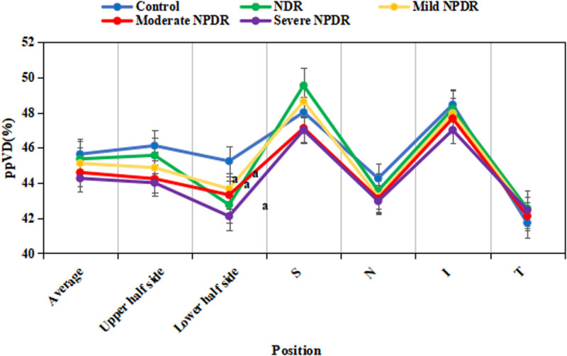
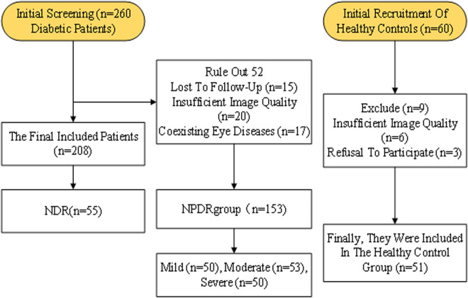
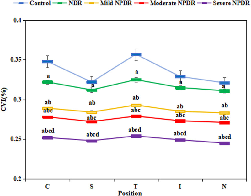
目的:探讨早期糖尿病视网膜病变(DR)的结构参数变化。材料与方法:本研究为回顾性分析,纳入南通大学附属第三医院2024年1月至2024年12月住院的早期DR患者。采用扫描源光学相干断层扫描血管造影(SS-OCTA)技术将参与者分为非DR组(NDR组)和非增长性DR组(NPDR组,包括轻度、中度和重度亚组)。采用单因素方差分析(ANOVA)和Kruskal-Wallis检验比较各组间参数差异。结果:共纳入糖尿病患者208例(NDR组55例,NPDR组153例),健康对照51例。结果显示,NPDR组FAZ面积显著大于对照组(CG)(平均差值:+0.38±0.10 mm2, 95% CI [0.25 ~ 0.51], P < 0.001),且与疾病严重程度呈正相关(趋势检验P < 0.001)。与CG相比,NDR组和NPDR各阶段的脉络膜血管指数(CVI)、乳头周围血管密度(ppVD)、乳头周围视网膜神经纤维层厚度(pRNFL)、视网膜浅层和深层血管复体血管密度(VD)、总灌注面积(PA)、小血管密度(SVD)、椎间盘面积、中央凹中心半径300µm范围内血管密度(FD300)、毛细血管丛血流密度(p)均显著降低。SS-OCTA可以有效监测结构参数的变化,是评估早期DR进展的有价值的工具。
Assessment of retinal and choroidal structural and microvascular changes in early diabetic retinopathy using swept-source optical coherence tomography angiography.
Objective: It was to assess changes in structural parameters in early diabetic retinopathy (DR).
Materials and methodologies: This study is a retrospective analysis that included patients with early DR admitted to the Affiliated Third Hospital of Nantong University from January 2024 to December 2024. The participants were divided into the non-DR group (NDR group) and the non-proliferative DR group (NPDR group, which included mild, moderate, and severe subgroups) using swept-source optical coherence tomography angiography (SS-OCTA) technology. One-way analysis of variance (ANOVA) and the Kruskal-Wallis test were used to compare parameter differences among the groups.
Results: A total of 208 diabetic patients were included (55 in the NDR group, 153 in the NPDR group) and 51 healthy controls. The results showed that the FAZ area in the NPDR group was significantly larger than that in the control group (CG) (mean difference: +0.38 ± 0.10 mm2, 95% CI [0.25-0.51], P < 0.001), and it was positively correlated with disease severity (trend test P < 0.001). Relative to the CG, NDR group and various stages of NPDR group exhibited greatly lower values in choroidal vascular index (CVI), peripapillary vascular density (ppVD), peripapillary retinal nerve fiber layer thickness (pRNFL), vascular density (VD) in both the superficial and deep retinal vascular complexes, total perfusion area (PA), small vessel density (SVD), disc area, vascular density (FD300) within a 300 µm radius of the foveal center, and capillary plexus blood flow density (P<0.05). NPDR group showed progressively lower values than NDR group, with severity increasing as the condition worsened (P<0.05).
Conclusion: SS-OCTA can effectively monitor changes in structural parameters and serves as a valuable tool for evaluating the progression of early DR.
期刊介绍:
PLOS ONE is an international, peer-reviewed, open-access, online publication. PLOS ONE welcomes reports on primary research from any scientific discipline. It provides:
* Open-access—freely accessible online, authors retain copyright
* Fast publication times
* Peer review by expert, practicing researchers
* Post-publication tools to indicate quality and impact
* Community-based dialogue on articles
* Worldwide media coverage

 求助内容:
求助内容: 应助结果提醒方式:


