Hannelore Ceuppens, Kirsten De Ridder, Thomas Ertveldt, Katty Zeven, Wout De Mey, Ana Rita Pombo Antunes, Laurent Navarro, Nina Dumauthioz, Tony Lahoutte, Jens M Debacker, Nick Devoogdt, Marleen Keyaerts, Matthias D'Huyvetter, Cleo Goyvaerts, Karine Breckpot
{"title":"临床前肿瘤模型中靶向成纤维细胞活化蛋白α的α和β放射性核素治疗后的免疫调节。","authors":"Hannelore Ceuppens, Kirsten De Ridder, Thomas Ertveldt, Katty Zeven, Wout De Mey, Ana Rita Pombo Antunes, Laurent Navarro, Nina Dumauthioz, Tony Lahoutte, Jens M Debacker, Nick Devoogdt, Marleen Keyaerts, Matthias D'Huyvetter, Cleo Goyvaerts, Karine Breckpot","doi":"10.1080/2162402X.2025.2540054","DOIUrl":null,"url":null,"abstract":"<p><p>α- and β<sup>-</sup>-emitting radionuclides targeting human fibroblast activation protein-α (hFAP) are under investigation for cancer therapy. In prior work, analysis of the tumor microenvironment 24 h after therapy completion indicated therapy-induced immune activation. Here, we analyzed systemic immune responses at varying timepoints during treatment to further elucidate the immune-stimulating effects of the therapy. Moreover, we analyzed end-stage tumors to gain insight in potential mechanisms of therapy resistance. Single domain antibody 4AH29 that binds hFAP was labeled with <sup>131</sup>I or <sup>225</sup>Ac, generating [<sup>131</sup>I]I-GMIB-4AH29 and [<sup>225</sup>Ac]Ac-DOTA-4AH29, respectively. These were used to treat C57BL/6 mice bearing subcutaneous TC-1-hFAP tumors. Blood analysis was conducted using flow cytometry, while tumor characterization was performed using flow cytometry and RNA sequencing. Given the distinct properties and doses of both radiopharmaceuticals, no head-to-head comparison was performed. Both treatments activated inflammatory responses in the tumor. Increased PD-1 expression on CD8<sup>+</sup> T-cells was observed following both treatments in the tumor and periphery. In the tumor, [<sup>131</sup>I]I-GMIB-4AH29 therapy uniquely induced the expression of genes involved in tumor cell replication, TNF-α, IL-6/STAT3, IL-2/STAT5 and complement pathways, while in the blood [<sup>131</sup>I]I-GMIB-4AH29 therapy upregulated SIRPα on monocytes and TIGIT on NK cells, and downregulated CD86 expression on monocytes. Longitudinal blood immune cell analysis showed changes in composition and phenotype early in therapy, e.g. in effector and regulatory T-cells. Overall, this study corroborates the immune sensitizing capacity of α- and β<sup>-</sup>-emitting radionuclides, triggering a variety of inflammatory effector responses.</p>","PeriodicalId":48714,"journal":{"name":"Oncoimmunology","volume":"14 1","pages":"2540054"},"PeriodicalIF":6.5000,"publicationDate":"2025-12-01","publicationTypes":"Journal Article","fieldsOfStudy":null,"isOpenAccess":false,"openAccessPdf":"https://www.ncbi.nlm.nih.gov/pmc/articles/PMC12320817/pdf/","citationCount":"0","resultStr":"{\"title\":\"Immune modulation following α and β<sup>-</sup> radionuclide therapy targeting fibroblast activation protein-α in a preclinical tumor model.\",\"authors\":\"Hannelore Ceuppens, Kirsten De Ridder, Thomas Ertveldt, Katty Zeven, Wout De Mey, Ana Rita Pombo Antunes, Laurent Navarro, Nina Dumauthioz, Tony Lahoutte, Jens M Debacker, Nick Devoogdt, Marleen Keyaerts, Matthias D'Huyvetter, Cleo Goyvaerts, Karine Breckpot\",\"doi\":\"10.1080/2162402X.2025.2540054\",\"DOIUrl\":null,\"url\":null,\"abstract\":\"<p><p>α- and β<sup>-</sup>-emitting radionuclides targeting human fibroblast activation protein-α (hFAP) are under investigation for cancer therapy. In prior work, analysis of the tumor microenvironment 24 h after therapy completion indicated therapy-induced immune activation. Here, we analyzed systemic immune responses at varying timepoints during treatment to further elucidate the immune-stimulating effects of the therapy. Moreover, we analyzed end-stage tumors to gain insight in potential mechanisms of therapy resistance. Single domain antibody 4AH29 that binds hFAP was labeled with <sup>131</sup>I or <sup>225</sup>Ac, generating [<sup>131</sup>I]I-GMIB-4AH29 and [<sup>225</sup>Ac]Ac-DOTA-4AH29, respectively. These were used to treat C57BL/6 mice bearing subcutaneous TC-1-hFAP tumors. Blood analysis was conducted using flow cytometry, while tumor characterization was performed using flow cytometry and RNA sequencing. Given the distinct properties and doses of both radiopharmaceuticals, no head-to-head comparison was performed. Both treatments activated inflammatory responses in the tumor. Increased PD-1 expression on CD8<sup>+</sup> T-cells was observed following both treatments in the tumor and periphery. In the tumor, [<sup>131</sup>I]I-GMIB-4AH29 therapy uniquely induced the expression of genes involved in tumor cell replication, TNF-α, IL-6/STAT3, IL-2/STAT5 and complement pathways, while in the blood [<sup>131</sup>I]I-GMIB-4AH29 therapy upregulated SIRPα on monocytes and TIGIT on NK cells, and downregulated CD86 expression on monocytes. Longitudinal blood immune cell analysis showed changes in composition and phenotype early in therapy, e.g. in effector and regulatory T-cells. Overall, this study corroborates the immune sensitizing capacity of α- and β<sup>-</sup>-emitting radionuclides, triggering a variety of inflammatory effector responses.</p>\",\"PeriodicalId\":48714,\"journal\":{\"name\":\"Oncoimmunology\",\"volume\":\"14 1\",\"pages\":\"2540054\"},\"PeriodicalIF\":6.5000,\"publicationDate\":\"2025-12-01\",\"publicationTypes\":\"Journal Article\",\"fieldsOfStudy\":null,\"isOpenAccess\":false,\"openAccessPdf\":\"https://www.ncbi.nlm.nih.gov/pmc/articles/PMC12320817/pdf/\",\"citationCount\":\"0\",\"resultStr\":null,\"platform\":\"Semanticscholar\",\"paperid\":null,\"PeriodicalName\":\"Oncoimmunology\",\"FirstCategoryId\":\"3\",\"ListUrlMain\":\"https://doi.org/10.1080/2162402X.2025.2540054\",\"RegionNum\":2,\"RegionCategory\":\"医学\",\"ArticlePicture\":[],\"TitleCN\":null,\"AbstractTextCN\":null,\"PMCID\":null,\"EPubDate\":\"2025/8/1 0:00:00\",\"PubModel\":\"Epub\",\"JCR\":\"Q1\",\"JCRName\":\"IMMUNOLOGY\",\"Score\":null,\"Total\":0}","platform":"Semanticscholar","paperid":null,"PeriodicalName":"Oncoimmunology","FirstCategoryId":"3","ListUrlMain":"https://doi.org/10.1080/2162402X.2025.2540054","RegionNum":2,"RegionCategory":"医学","ArticlePicture":[],"TitleCN":null,"AbstractTextCN":null,"PMCID":null,"EPubDate":"2025/8/1 0:00:00","PubModel":"Epub","JCR":"Q1","JCRName":"IMMUNOLOGY","Score":null,"Total":0}
引用次数: 0
摘要
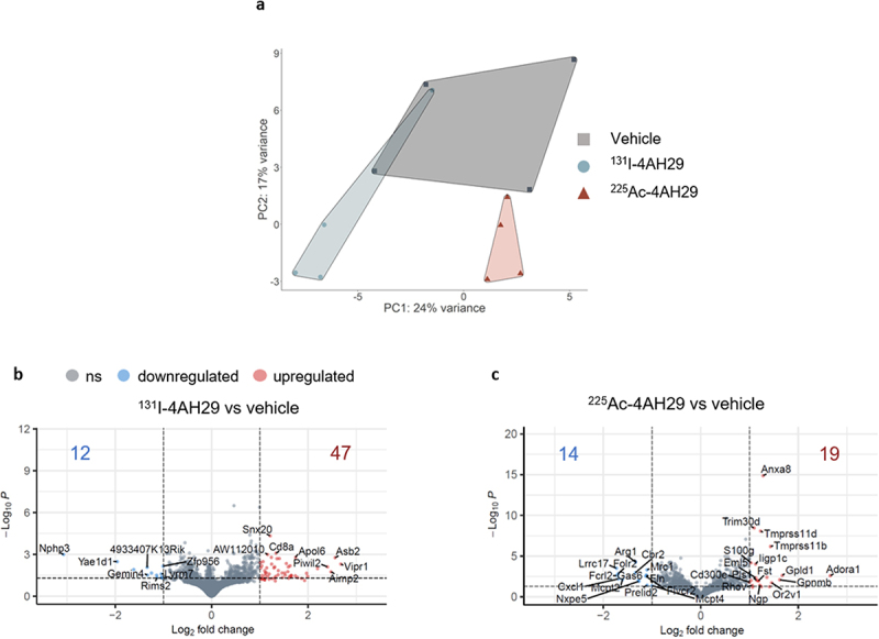
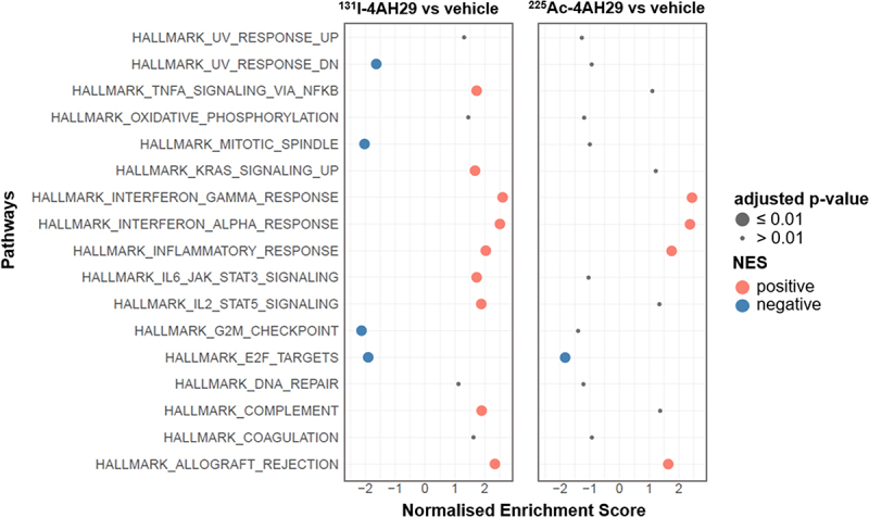
靶向人成纤维细胞活化蛋白-α (hFAP)的α-和β-放射核素正在研究用于癌症治疗。在之前的研究中,在治疗结束后24小时对肿瘤微环境的分析表明,治疗诱导了免疫激活。在这里,我们分析了治疗过程中不同时间点的全身免疫反应,以进一步阐明治疗的免疫刺激作用。此外,我们分析了终末期肿瘤,以深入了解治疗耐药的潜在机制。结合hFAP的单域抗体4AH29分别用131I或225Ac标记,分别生成[131I]I-GMIB-4AH29和[225Ac]Ac-DOTA-4AH29。这些药物用于治疗皮下TC-1-hFAP肿瘤的C57BL/6小鼠。采用流式细胞术进行血液分析,采用流式细胞术和RNA测序进行肿瘤表征。鉴于这两种放射性药物的特性和剂量不同,没有进行正面比较。两种治疗方法都激活了肿瘤的炎症反应。在肿瘤和外周治疗后,PD-1在CD8+ t细胞上的表达均升高。在肿瘤中,[131I] i - gmb - 4ah29治疗独特地诱导肿瘤细胞复制相关基因、TNF-α、IL-6/STAT3、IL-2/STAT5和补体通路的表达,而在血液中[131I] i - gmb - 4ah29治疗上调单核细胞上的SIRPα和NK细胞上的TIGIT,下调单核细胞上CD86的表达。纵向血液免疫细胞分析显示,在治疗早期,效应t细胞和调节性t细胞的组成和表型发生了变化。总的来说,本研究证实了α-和β-发射放射性核素的免疫增敏能力,引发各种炎症效应反应。
Immune modulation following α and β- radionuclide therapy targeting fibroblast activation protein-α in a preclinical tumor model.
α- and β--emitting radionuclides targeting human fibroblast activation protein-α (hFAP) are under investigation for cancer therapy. In prior work, analysis of the tumor microenvironment 24 h after therapy completion indicated therapy-induced immune activation. Here, we analyzed systemic immune responses at varying timepoints during treatment to further elucidate the immune-stimulating effects of the therapy. Moreover, we analyzed end-stage tumors to gain insight in potential mechanisms of therapy resistance. Single domain antibody 4AH29 that binds hFAP was labeled with 131I or 225Ac, generating [131I]I-GMIB-4AH29 and [225Ac]Ac-DOTA-4AH29, respectively. These were used to treat C57BL/6 mice bearing subcutaneous TC-1-hFAP tumors. Blood analysis was conducted using flow cytometry, while tumor characterization was performed using flow cytometry and RNA sequencing. Given the distinct properties and doses of both radiopharmaceuticals, no head-to-head comparison was performed. Both treatments activated inflammatory responses in the tumor. Increased PD-1 expression on CD8+ T-cells was observed following both treatments in the tumor and periphery. In the tumor, [131I]I-GMIB-4AH29 therapy uniquely induced the expression of genes involved in tumor cell replication, TNF-α, IL-6/STAT3, IL-2/STAT5 and complement pathways, while in the blood [131I]I-GMIB-4AH29 therapy upregulated SIRPα on monocytes and TIGIT on NK cells, and downregulated CD86 expression on monocytes. Longitudinal blood immune cell analysis showed changes in composition and phenotype early in therapy, e.g. in effector and regulatory T-cells. Overall, this study corroborates the immune sensitizing capacity of α- and β--emitting radionuclides, triggering a variety of inflammatory effector responses.
期刊介绍:
OncoImmunology is a dynamic, high-profile, open access journal that comprehensively covers tumor immunology and immunotherapy.
As cancer immunotherapy advances, OncoImmunology is committed to publishing top-tier research encompassing all facets of basic and applied tumor immunology.
The journal covers a wide range of topics, including:
-Basic and translational studies in immunology of both solid and hematological malignancies
-Inflammation, innate and acquired immune responses against cancer
-Mechanisms of cancer immunoediting and immune evasion
-Modern immunotherapies, including immunomodulators, immune checkpoint inhibitors, T-cell, NK-cell, and macrophage engagers, and CAR T cells
-Immunological effects of conventional anticancer therapies.

 求助内容:
求助内容: 应助结果提醒方式:


