{"title":"分化型甲状腺癌术后碘-131全身显像肝脏积聚定量评估疾病进展预测因子","authors":"Michihiro Nakayama, Kenta Nomura, Sho Kamieda, Ippei Yoshida, Atsushi Fujiya, Takahiro Uno, Atsutaka Okizaki","doi":"10.4274/mirt.galenos.2025.71542","DOIUrl":null,"url":null,"abstract":"<p><strong>Objectives: </strong>A Iodine-131 (<sup>131</sup>I) whole body scan (WBS) is performed to evaluate the treatment response after radioactive iodine (RAI) therapy. Despite the clinical relevance of RAI-refractory differentiated thyroid cancer, a consensus on its precise definition remains lacking. This study investigates the potential utility of hepatic <sup>131</sup>I accumulation as an early predictor for tumor recurrence or progression after RAI administration.</p><p><strong>Methods: </strong>Of 814 patients receiving care at our institution, we enrolled 225 patients who exhibited no accumulation of RAI in the remnant tissues or other lesions on <sup>131</sup>I WBS. We quantified the hepatic uptake ratio [defined as (hepatic uptake/background uptake (H/B)] from WBS. All patients were categorized into group A (H/B ≤1.5) and group B (H/B >1.5), and we assessed between-group differences. The Kaplan-Meier method and Log-rank test were used to analyze the progression-free survival (PFS). Using the Cox proportional hazards model, we identified independent prognostic factors from among the seven known prognostic factors, i.e., H/B, thyroglobulin, sex, age, stage, total <sup>131</sup>I dose, and final therapeutic dose.</p><p><strong>Results: </strong>The 5-year and median PFS were 98.8% and 114.7 months in group A (n: 171) compared with 24.1% and 42.7% months in group B (n: 54), respectively. Group B showed a significant correlation with poor prognosis (p<0.00001). Of the seven prognostic factors, H/B exhibited the highest impact on patient outcomes (hazards ratio for recurrence/disease progression, 42.156; 95% confidence interval: 8.750-203.106).</p><p><strong>Conclusion: </strong>Quantitative evaluation of hepatic uptake on <sup>131</sup>I WBS provides a marker that may help identify patients with differentiated thyroid cancer who are at a high risk of disease progression/recurrence immediately after RAI therapy.</p>","PeriodicalId":44681,"journal":{"name":"Molecular Imaging and Radionuclide Therapy","volume":" ","pages":"194-201"},"PeriodicalIF":1.1000,"publicationDate":"2025-10-08","publicationTypes":"Journal Article","fieldsOfStudy":null,"isOpenAccess":false,"openAccessPdf":"https://www.ncbi.nlm.nih.gov/pmc/articles/PMC12505212/pdf/","citationCount":"0","resultStr":"{\"title\":\"A Disease Progression Predictor by Quantitative Assessment of the Hepatic Accumulation on Postablative Iodine-131 Whole-Body Image in Differentiated Thyroid Cancer.\",\"authors\":\"Michihiro Nakayama, Kenta Nomura, Sho Kamieda, Ippei Yoshida, Atsushi Fujiya, Takahiro Uno, Atsutaka Okizaki\",\"doi\":\"10.4274/mirt.galenos.2025.71542\",\"DOIUrl\":null,\"url\":null,\"abstract\":\"<p><strong>Objectives: </strong>A Iodine-131 (<sup>131</sup>I) whole body scan (WBS) is performed to evaluate the treatment response after radioactive iodine (RAI) therapy. Despite the clinical relevance of RAI-refractory differentiated thyroid cancer, a consensus on its precise definition remains lacking. This study investigates the potential utility of hepatic <sup>131</sup>I accumulation as an early predictor for tumor recurrence or progression after RAI administration.</p><p><strong>Methods: </strong>Of 814 patients receiving care at our institution, we enrolled 225 patients who exhibited no accumulation of RAI in the remnant tissues or other lesions on <sup>131</sup>I WBS. We quantified the hepatic uptake ratio [defined as (hepatic uptake/background uptake (H/B)] from WBS. All patients were categorized into group A (H/B ≤1.5) and group B (H/B >1.5), and we assessed between-group differences. The Kaplan-Meier method and Log-rank test were used to analyze the progression-free survival (PFS). Using the Cox proportional hazards model, we identified independent prognostic factors from among the seven known prognostic factors, i.e., H/B, thyroglobulin, sex, age, stage, total <sup>131</sup>I dose, and final therapeutic dose.</p><p><strong>Results: </strong>The 5-year and median PFS were 98.8% and 114.7 months in group A (n: 171) compared with 24.1% and 42.7% months in group B (n: 54), respectively. Group B showed a significant correlation with poor prognosis (p<0.00001). Of the seven prognostic factors, H/B exhibited the highest impact on patient outcomes (hazards ratio for recurrence/disease progression, 42.156; 95% confidence interval: 8.750-203.106).</p><p><strong>Conclusion: </strong>Quantitative evaluation of hepatic uptake on <sup>131</sup>I WBS provides a marker that may help identify patients with differentiated thyroid cancer who are at a high risk of disease progression/recurrence immediately after RAI therapy.</p>\",\"PeriodicalId\":44681,\"journal\":{\"name\":\"Molecular Imaging and Radionuclide Therapy\",\"volume\":\" \",\"pages\":\"194-201\"},\"PeriodicalIF\":1.1000,\"publicationDate\":\"2025-10-08\",\"publicationTypes\":\"Journal Article\",\"fieldsOfStudy\":null,\"isOpenAccess\":false,\"openAccessPdf\":\"https://www.ncbi.nlm.nih.gov/pmc/articles/PMC12505212/pdf/\",\"citationCount\":\"0\",\"resultStr\":null,\"platform\":\"Semanticscholar\",\"paperid\":null,\"PeriodicalName\":\"Molecular Imaging and Radionuclide Therapy\",\"FirstCategoryId\":\"1085\",\"ListUrlMain\":\"https://doi.org/10.4274/mirt.galenos.2025.71542\",\"RegionNum\":0,\"RegionCategory\":null,\"ArticlePicture\":[],\"TitleCN\":null,\"AbstractTextCN\":null,\"PMCID\":null,\"EPubDate\":\"2025/8/1 0:00:00\",\"PubModel\":\"Epub\",\"JCR\":\"Q4\",\"JCRName\":\"RADIOLOGY, NUCLEAR MEDICINE & MEDICAL IMAGING\",\"Score\":null,\"Total\":0}","platform":"Semanticscholar","paperid":null,"PeriodicalName":"Molecular Imaging and Radionuclide Therapy","FirstCategoryId":"1085","ListUrlMain":"https://doi.org/10.4274/mirt.galenos.2025.71542","RegionNum":0,"RegionCategory":null,"ArticlePicture":[],"TitleCN":null,"AbstractTextCN":null,"PMCID":null,"EPubDate":"2025/8/1 0:00:00","PubModel":"Epub","JCR":"Q4","JCRName":"RADIOLOGY, NUCLEAR MEDICINE & MEDICAL IMAGING","Score":null,"Total":0}
A Disease Progression Predictor by Quantitative Assessment of the Hepatic Accumulation on Postablative Iodine-131 Whole-Body Image in Differentiated Thyroid Cancer.
Objectives: A Iodine-131 (131I) whole body scan (WBS) is performed to evaluate the treatment response after radioactive iodine (RAI) therapy. Despite the clinical relevance of RAI-refractory differentiated thyroid cancer, a consensus on its precise definition remains lacking. This study investigates the potential utility of hepatic 131I accumulation as an early predictor for tumor recurrence or progression after RAI administration.
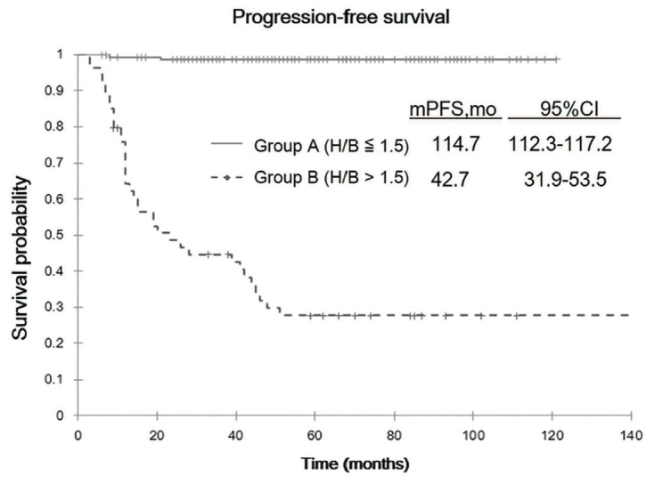
Methods: Of 814 patients receiving care at our institution, we enrolled 225 patients who exhibited no accumulation of RAI in the remnant tissues or other lesions on 131I WBS. We quantified the hepatic uptake ratio [defined as (hepatic uptake/background uptake (H/B)] from WBS. All patients were categorized into group A (H/B ≤1.5) and group B (H/B >1.5), and we assessed between-group differences. The Kaplan-Meier method and Log-rank test were used to analyze the progression-free survival (PFS). Using the Cox proportional hazards model, we identified independent prognostic factors from among the seven known prognostic factors, i.e., H/B, thyroglobulin, sex, age, stage, total 131I dose, and final therapeutic dose.
Results: The 5-year and median PFS were 98.8% and 114.7 months in group A (n: 171) compared with 24.1% and 42.7% months in group B (n: 54), respectively. Group B showed a significant correlation with poor prognosis (p<0.00001). Of the seven prognostic factors, H/B exhibited the highest impact on patient outcomes (hazards ratio for recurrence/disease progression, 42.156; 95% confidence interval: 8.750-203.106).
Conclusion: Quantitative evaluation of hepatic uptake on 131I WBS provides a marker that may help identify patients with differentiated thyroid cancer who are at a high risk of disease progression/recurrence immediately after RAI therapy.
期刊介绍:
Molecular Imaging and Radionuclide Therapy (Mol Imaging Radionucl Ther, MIRT) is publishes original research articles, invited reviews, editorials, short communications, letters, consensus statements, guidelines and case reports with a literature review on the topic, in the field of molecular imaging, multimodality imaging, nuclear medicine, radionuclide therapy, radiopharmacy, medical physics, dosimetry and radiobiology.

 求助内容:
求助内容: 应助结果提醒方式:


