{"title":"扫描源OCT血管造影衍生的健康人群乳头周围血管密度区域规范数据。","authors":"Zhuoyan Yang, Ruihua Jing, Yidan Wu, Jiaxin Gao, Shiyuan Hu, Jianming Wang","doi":"10.1167/tvst.14.8.5","DOIUrl":null,"url":null,"abstract":"<p><strong>Purpose: </strong>The purpose of this study was to investigate the distribution of peripapillary vessel density (VD) across different regions of retinal and choroidal layers and analyze its influencing factors after axial length (AL) correction.</p><p><strong>Methods: </strong>This study included 337 eyes without significant fundus abnormalities or systemic conditions affecting blood flow. Swept-source optical coherence tomography angiography (SS-OCTA) was utilized to measure peripapillary VD and associated ocular parameters.</p><p><strong>Results: </strong>VD in all regions of the choroidal layer decreased significantly with increasing AL among AL groups. In non-highly myopic eyes, VD followed the pattern temporal (T) > superior nasal (SN) > inferior nasal (IN) in the inner retina and superficial vascular complex (SVC), whereas in the deep vascular complex (DVC), it was inferior (I) > superior (S). In the choroid, VD ranked as nasal (N) > T. Highly myopic eyes showed higher temporal but lower nasal VD in the inner retina and SVC, whereas DVC exhibited the opposite trend. Choroidal VD decreased significantly across all regions, most prominently in the T hemisphere. Multivariable analysis identified age, signal strength, and retinal nerve fiber layer (RNFL) thickness as key determinants of inner retinal VD, whereas AL, signal strength, vertical cup-to-disc ratio (C/D), Bruch's membrane opening (BMO), and optic disc-fovea distance (DFD) significantly influenced choroidal VD.</p><p><strong>Conclusions: </strong>Inner retinal VD followed T > SN > IN, primarily contributed by the SVC, whereas choroidal VD followed N > T. With increasing AL, choroidal VD declined across all regions, most prominently in the T hemisphere, whereas the inner retinal VD trends varied by region and layer.</p><p><strong>Translational relevance: </strong>The findings of this study may contribute to the early warning of the disease and provide an important theoretical basis for the study of myopia-related microcirculatory alteration mechanisms.</p>","PeriodicalId":23322,"journal":{"name":"Translational Vision Science & Technology","volume":"14 8","pages":"5"},"PeriodicalIF":2.6000,"publicationDate":"2025-08-01","publicationTypes":"Journal Article","fieldsOfStudy":null,"isOpenAccess":false,"openAccessPdf":"https://www.ncbi.nlm.nih.gov/pmc/articles/PMC12320905/pdf/","citationCount":"0","resultStr":"{\"title\":\"Swept-Source OCT Angiography-Derived Regional Normative Data of Peripapillary Vessel Density in Healthy Populations.\",\"authors\":\"Zhuoyan Yang, Ruihua Jing, Yidan Wu, Jiaxin Gao, Shiyuan Hu, Jianming Wang\",\"doi\":\"10.1167/tvst.14.8.5\",\"DOIUrl\":null,\"url\":null,\"abstract\":\"<p><strong>Purpose: </strong>The purpose of this study was to investigate the distribution of peripapillary vessel density (VD) across different regions of retinal and choroidal layers and analyze its influencing factors after axial length (AL) correction.</p><p><strong>Methods: </strong>This study included 337 eyes without significant fundus abnormalities or systemic conditions affecting blood flow. Swept-source optical coherence tomography angiography (SS-OCTA) was utilized to measure peripapillary VD and associated ocular parameters.</p><p><strong>Results: </strong>VD in all regions of the choroidal layer decreased significantly with increasing AL among AL groups. In non-highly myopic eyes, VD followed the pattern temporal (T) > superior nasal (SN) > inferior nasal (IN) in the inner retina and superficial vascular complex (SVC), whereas in the deep vascular complex (DVC), it was inferior (I) > superior (S). In the choroid, VD ranked as nasal (N) > T. Highly myopic eyes showed higher temporal but lower nasal VD in the inner retina and SVC, whereas DVC exhibited the opposite trend. Choroidal VD decreased significantly across all regions, most prominently in the T hemisphere. Multivariable analysis identified age, signal strength, and retinal nerve fiber layer (RNFL) thickness as key determinants of inner retinal VD, whereas AL, signal strength, vertical cup-to-disc ratio (C/D), Bruch's membrane opening (BMO), and optic disc-fovea distance (DFD) significantly influenced choroidal VD.</p><p><strong>Conclusions: </strong>Inner retinal VD followed T > SN > IN, primarily contributed by the SVC, whereas choroidal VD followed N > T. With increasing AL, choroidal VD declined across all regions, most prominently in the T hemisphere, whereas the inner retinal VD trends varied by region and layer.</p><p><strong>Translational relevance: </strong>The findings of this study may contribute to the early warning of the disease and provide an important theoretical basis for the study of myopia-related microcirculatory alteration mechanisms.</p>\",\"PeriodicalId\":23322,\"journal\":{\"name\":\"Translational Vision Science & Technology\",\"volume\":\"14 8\",\"pages\":\"5\"},\"PeriodicalIF\":2.6000,\"publicationDate\":\"2025-08-01\",\"publicationTypes\":\"Journal Article\",\"fieldsOfStudy\":null,\"isOpenAccess\":false,\"openAccessPdf\":\"https://www.ncbi.nlm.nih.gov/pmc/articles/PMC12320905/pdf/\",\"citationCount\":\"0\",\"resultStr\":null,\"platform\":\"Semanticscholar\",\"paperid\":null,\"PeriodicalName\":\"Translational Vision Science & Technology\",\"FirstCategoryId\":\"3\",\"ListUrlMain\":\"https://doi.org/10.1167/tvst.14.8.5\",\"RegionNum\":3,\"RegionCategory\":\"医学\",\"ArticlePicture\":[],\"TitleCN\":null,\"AbstractTextCN\":null,\"PMCID\":null,\"EPubDate\":\"\",\"PubModel\":\"\",\"JCR\":\"Q2\",\"JCRName\":\"OPHTHALMOLOGY\",\"Score\":null,\"Total\":0}","platform":"Semanticscholar","paperid":null,"PeriodicalName":"Translational Vision Science & Technology","FirstCategoryId":"3","ListUrlMain":"https://doi.org/10.1167/tvst.14.8.5","RegionNum":3,"RegionCategory":"医学","ArticlePicture":[],"TitleCN":null,"AbstractTextCN":null,"PMCID":null,"EPubDate":"","PubModel":"","JCR":"Q2","JCRName":"OPHTHALMOLOGY","Score":null,"Total":0}
Swept-Source OCT Angiography-Derived Regional Normative Data of Peripapillary Vessel Density in Healthy Populations.
Purpose: The purpose of this study was to investigate the distribution of peripapillary vessel density (VD) across different regions of retinal and choroidal layers and analyze its influencing factors after axial length (AL) correction.
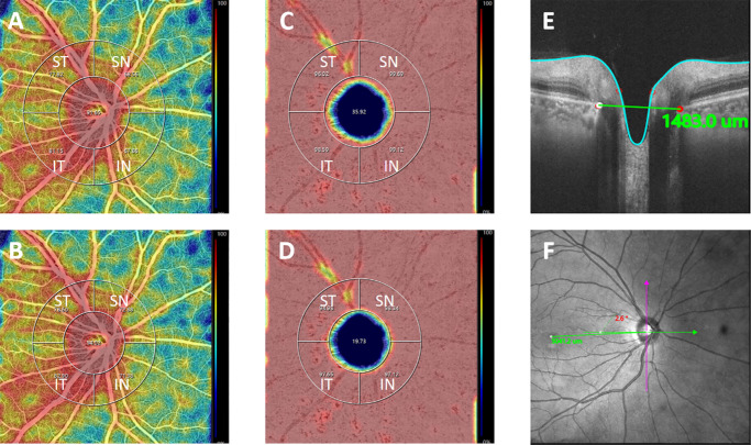
Methods: This study included 337 eyes without significant fundus abnormalities or systemic conditions affecting blood flow. Swept-source optical coherence tomography angiography (SS-OCTA) was utilized to measure peripapillary VD and associated ocular parameters.
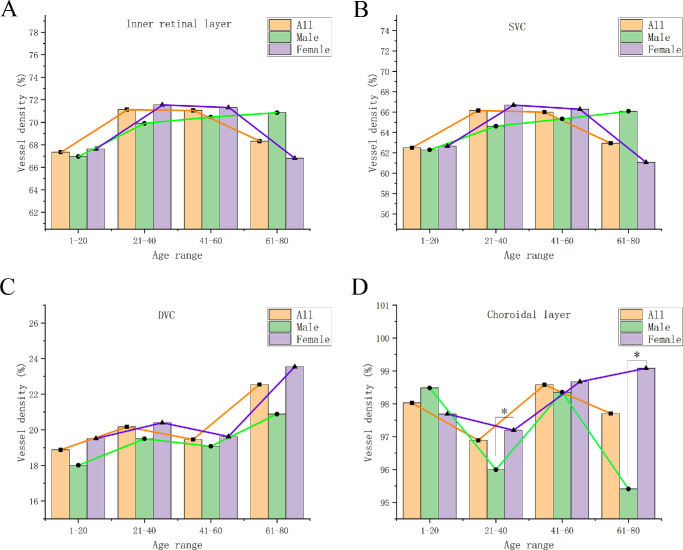
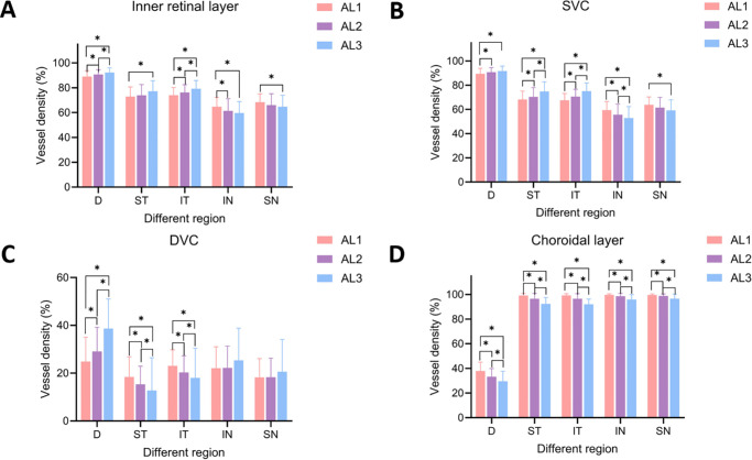
Results: VD in all regions of the choroidal layer decreased significantly with increasing AL among AL groups. In non-highly myopic eyes, VD followed the pattern temporal (T) > superior nasal (SN) > inferior nasal (IN) in the inner retina and superficial vascular complex (SVC), whereas in the deep vascular complex (DVC), it was inferior (I) > superior (S). In the choroid, VD ranked as nasal (N) > T. Highly myopic eyes showed higher temporal but lower nasal VD in the inner retina and SVC, whereas DVC exhibited the opposite trend. Choroidal VD decreased significantly across all regions, most prominently in the T hemisphere. Multivariable analysis identified age, signal strength, and retinal nerve fiber layer (RNFL) thickness as key determinants of inner retinal VD, whereas AL, signal strength, vertical cup-to-disc ratio (C/D), Bruch's membrane opening (BMO), and optic disc-fovea distance (DFD) significantly influenced choroidal VD.
Conclusions: Inner retinal VD followed T > SN > IN, primarily contributed by the SVC, whereas choroidal VD followed N > T. With increasing AL, choroidal VD declined across all regions, most prominently in the T hemisphere, whereas the inner retinal VD trends varied by region and layer.
Translational relevance: The findings of this study may contribute to the early warning of the disease and provide an important theoretical basis for the study of myopia-related microcirculatory alteration mechanisms.
期刊介绍:
Translational Vision Science & Technology (TVST), an official journal of the Association for Research in Vision and Ophthalmology (ARVO), an international organization whose purpose is to advance research worldwide into understanding the visual system and preventing, treating and curing its disorders, is an online, open access, peer-reviewed journal emphasizing multidisciplinary research that bridges the gap between basic research and clinical care. A highly qualified and diverse group of Associate Editors and Editorial Board Members is led by Editor-in-Chief Marco Zarbin, MD, PhD, FARVO.
The journal covers a broad spectrum of work, including but not limited to:
Applications of stem cell technology for regenerative medicine,
Development of new animal models of human diseases,
Tissue bioengineering,
Chemical engineering to improve virus-based gene delivery,
Nanotechnology for drug delivery,
Design and synthesis of artificial extracellular matrices,
Development of a true microsurgical operating environment,
Refining data analysis algorithms to improve in vivo imaging technology,
Results of Phase 1 clinical trials,
Reverse translational ("bedside to bench") research.
TVST seeks manuscripts from scientists and clinicians with diverse backgrounds ranging from basic chemistry to ophthalmic surgery that will advance or change the way we understand and/or treat vision-threatening diseases. TVST encourages the use of color, multimedia, hyperlinks, program code and other digital enhancements.

 求助内容:
求助内容: 应助结果提醒方式:


