{"title":"食管保留囊肿伴高氟脱氧葡萄糖摄取PET/CT 1例。","authors":"Byonggu An, Hiroshi Yamamoto, Yasumitsu Oe, Takeshi Togawa, Kazumi Shimamoto, Hiromitsu Ban, Tetsuya Abe, Yuki Morimoto, Takashi Matsunaga, Toru Imagami, Akira Sogawa, Nobuyuki Takao, Shizuki Takemura, Akiyoshi Mizumoto","doi":"10.70352/scrj.cr.25-0348","DOIUrl":null,"url":null,"abstract":"<p><strong>Introduction: </strong>Esophageal retention cysts are rare, benign lesions that can mimic submucosal tumors. Their clinical presentation and imaging characteristics may lead to diagnostic challenges, particularly when fluorodeoxyglucose-positron emission tomography/CT (FDG-PET/CT) shows increased uptake, raising suspicion of malignancy.</p><p><strong>Case presentation: </strong>A 77-year-old man presented with epigastric pain. Upper gastrointestinal endoscopy revealed an esophageal mass, prompting referral to our hospital. Endoscopic ultrasonography (EUS) identified a hypoechoic submucosal tumor with multiple cystic components in the lower esophagus. However, EUS-guided fine-needle aspiration (EUS-FNA) did not yield a definitive diagnosis. CT scan demonstrated a 60-mm space-occupying lesion (SOL) in the lower thoracic esophagus with peripheral contrast enhancement and a central low-density area. MRI revealed a SOL in the lower esophagus with high signal intensity on T2-weighted images and moderate signal intensity on T1-weighted images. The lesion contained cystic components exhibiting high T2 and low T1 signal intensities. FDG-PET/CT revealed intense FDG uptake, increasing from maximum standardized uptake value (SUVmax) 11 to 18 over time. Given the large size of the tumor, symptomatology, and inability to exclude malignancy-particularly high-risk gastrointestinal stromal tumor-surgical resection was performed. Laparoscopic esophagectomy was conducted using intraoperative endoscopy for tumor identification. The esophagus was transected proximally using a linear stapler, followed by extracorporeal gastric conduit reconstruction and the overlap technique was used to perform an esophagogastric anastomosis. Postoperatively, anastomotic leakage was detected on day 3, requiring emergency reoperation. The leak had resolved by POD 26, and the patient was discharged on day 48 after the second surgery (day 51 after the initial surgery). Histopathological examination revealed multiple cysts of varying sizes within the lamina propria, lined by columnar epithelium, with no evidence of malignancy. The final diagnosis was esophageal retention cyst.</p><p><strong>Conclusions: </strong>This case highlights the diagnostic challenge of esophageal retention cysts with high FDG uptake. While PET/CT is essential in oncologic imaging, FDG accumulation does not always indicate malignancy.</p>","PeriodicalId":22096,"journal":{"name":"Surgical Case Reports","volume":"11 1","pages":""},"PeriodicalIF":0.7000,"publicationDate":"2025-01-01","publicationTypes":"Journal Article","fieldsOfStudy":null,"isOpenAccess":false,"openAccessPdf":"https://www.ncbi.nlm.nih.gov/pmc/articles/PMC12313353/pdf/","citationCount":"0","resultStr":"{\"title\":\"A Case of Esophageal Retention Cyst with High Fluorodeoxyglucose Uptake on PET/CT Scan.\",\"authors\":\"Byonggu An, Hiroshi Yamamoto, Yasumitsu Oe, Takeshi Togawa, Kazumi Shimamoto, Hiromitsu Ban, Tetsuya Abe, Yuki Morimoto, Takashi Matsunaga, Toru Imagami, Akira Sogawa, Nobuyuki Takao, Shizuki Takemura, Akiyoshi Mizumoto\",\"doi\":\"10.70352/scrj.cr.25-0348\",\"DOIUrl\":null,\"url\":null,\"abstract\":\"<p><strong>Introduction: </strong>Esophageal retention cysts are rare, benign lesions that can mimic submucosal tumors. Their clinical presentation and imaging characteristics may lead to diagnostic challenges, particularly when fluorodeoxyglucose-positron emission tomography/CT (FDG-PET/CT) shows increased uptake, raising suspicion of malignancy.</p><p><strong>Case presentation: </strong>A 77-year-old man presented with epigastric pain. Upper gastrointestinal endoscopy revealed an esophageal mass, prompting referral to our hospital. Endoscopic ultrasonography (EUS) identified a hypoechoic submucosal tumor with multiple cystic components in the lower esophagus. However, EUS-guided fine-needle aspiration (EUS-FNA) did not yield a definitive diagnosis. CT scan demonstrated a 60-mm space-occupying lesion (SOL) in the lower thoracic esophagus with peripheral contrast enhancement and a central low-density area. MRI revealed a SOL in the lower esophagus with high signal intensity on T2-weighted images and moderate signal intensity on T1-weighted images. The lesion contained cystic components exhibiting high T2 and low T1 signal intensities. FDG-PET/CT revealed intense FDG uptake, increasing from maximum standardized uptake value (SUVmax) 11 to 18 over time. Given the large size of the tumor, symptomatology, and inability to exclude malignancy-particularly high-risk gastrointestinal stromal tumor-surgical resection was performed. Laparoscopic esophagectomy was conducted using intraoperative endoscopy for tumor identification. The esophagus was transected proximally using a linear stapler, followed by extracorporeal gastric conduit reconstruction and the overlap technique was used to perform an esophagogastric anastomosis. Postoperatively, anastomotic leakage was detected on day 3, requiring emergency reoperation. The leak had resolved by POD 26, and the patient was discharged on day 48 after the second surgery (day 51 after the initial surgery). Histopathological examination revealed multiple cysts of varying sizes within the lamina propria, lined by columnar epithelium, with no evidence of malignancy. The final diagnosis was esophageal retention cyst.</p><p><strong>Conclusions: </strong>This case highlights the diagnostic challenge of esophageal retention cysts with high FDG uptake. While PET/CT is essential in oncologic imaging, FDG accumulation does not always indicate malignancy.</p>\",\"PeriodicalId\":22096,\"journal\":{\"name\":\"Surgical Case Reports\",\"volume\":\"11 1\",\"pages\":\"\"},\"PeriodicalIF\":0.7000,\"publicationDate\":\"2025-01-01\",\"publicationTypes\":\"Journal Article\",\"fieldsOfStudy\":null,\"isOpenAccess\":false,\"openAccessPdf\":\"https://www.ncbi.nlm.nih.gov/pmc/articles/PMC12313353/pdf/\",\"citationCount\":\"0\",\"resultStr\":null,\"platform\":\"Semanticscholar\",\"paperid\":null,\"PeriodicalName\":\"Surgical Case Reports\",\"FirstCategoryId\":\"1085\",\"ListUrlMain\":\"https://doi.org/10.70352/scrj.cr.25-0348\",\"RegionNum\":0,\"RegionCategory\":null,\"ArticlePicture\":[],\"TitleCN\":null,\"AbstractTextCN\":null,\"PMCID\":null,\"EPubDate\":\"2025/7/29 0:00:00\",\"PubModel\":\"Epub\",\"JCR\":\"Q4\",\"JCRName\":\"SURGERY\",\"Score\":null,\"Total\":0}","platform":"Semanticscholar","paperid":null,"PeriodicalName":"Surgical Case Reports","FirstCategoryId":"1085","ListUrlMain":"https://doi.org/10.70352/scrj.cr.25-0348","RegionNum":0,"RegionCategory":null,"ArticlePicture":[],"TitleCN":null,"AbstractTextCN":null,"PMCID":null,"EPubDate":"2025/7/29 0:00:00","PubModel":"Epub","JCR":"Q4","JCRName":"SURGERY","Score":null,"Total":0}
引用次数: 0
摘要
简介:食管潴留囊肿是一种罕见的良性病变,可以模拟粘膜下肿瘤。它们的临床表现和影像学特征可能导致诊断困难,特别是当氟脱氧葡萄糖-正电子发射断层扫描/CT (FDG-PET/CT)显示摄取增加时,增加了对恶性肿瘤的怀疑。病例介绍:一名77岁男性,主诉胃脘痛。上消化道内窥镜检查发现食管肿块,提示转介至我院。内镜超声检查(EUS)发现食管下段有多发性囊性成分的低回声粘膜下肿瘤。然而,eus引导的细针抽吸(EUS-FNA)并没有产生明确的诊断。CT扫描显示下胸食道60毫米占位性病变(SOL),周围增强,中心低密度区。MRI示食管下段一SOL, t2加权高信号强度,t1加权中等信号强度。病变包含囊性成分,表现为高T2和低T1信号强度。FDG- pet /CT显示FDG摄取强烈,随时间从最大标准化摄取值(SUVmax) 11增加到18。考虑到肿瘤的大小、症状和无法排除恶性肿瘤,特别是高危胃肠道间质瘤,我们进行了手术切除。腹腔镜食管切除术采用术中内镜检查肿瘤。用线性吻合器近端切开食管,然后进行体外胃管重建,并使用重叠技术进行食管胃吻合。术后第3天发现吻合口漏,需要紧急再手术。经POD 26解决漏出,患者于第二次手术后第48天(首次手术后第51天)出院。组织病理学检查显示固有层内有多个大小不等的囊肿,内衬柱状上皮,无恶性肿瘤迹象。最终诊断为食道潴留囊肿。结论:本病例强调了食道保留囊肿伴高FDG摄取的诊断挑战。虽然PET/CT在肿瘤成像中是必不可少的,但FDG积聚并不总是表明恶性肿瘤。
A Case of Esophageal Retention Cyst with High Fluorodeoxyglucose Uptake on PET/CT Scan.
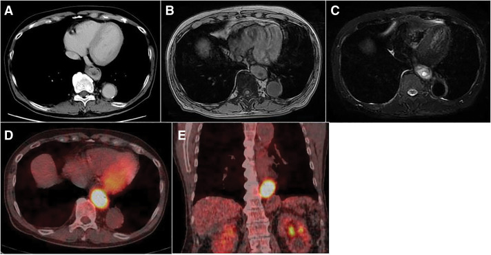
Introduction: Esophageal retention cysts are rare, benign lesions that can mimic submucosal tumors. Their clinical presentation and imaging characteristics may lead to diagnostic challenges, particularly when fluorodeoxyglucose-positron emission tomography/CT (FDG-PET/CT) shows increased uptake, raising suspicion of malignancy.
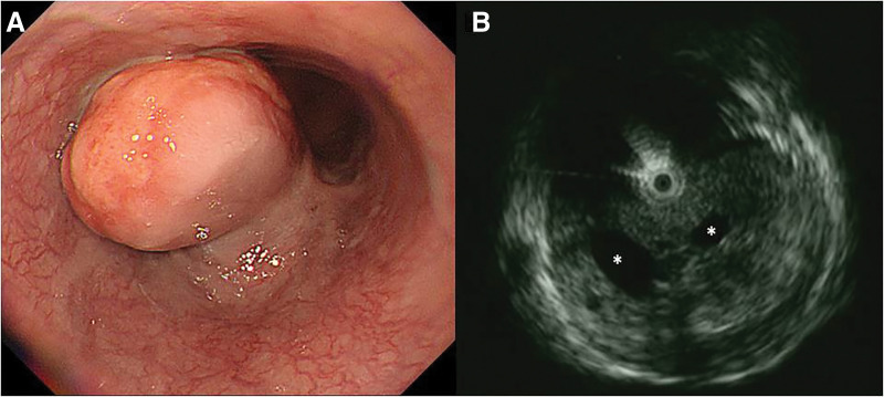
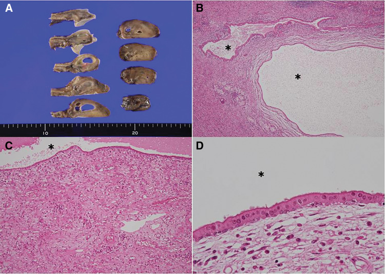
Case presentation: A 77-year-old man presented with epigastric pain. Upper gastrointestinal endoscopy revealed an esophageal mass, prompting referral to our hospital. Endoscopic ultrasonography (EUS) identified a hypoechoic submucosal tumor with multiple cystic components in the lower esophagus. However, EUS-guided fine-needle aspiration (EUS-FNA) did not yield a definitive diagnosis. CT scan demonstrated a 60-mm space-occupying lesion (SOL) in the lower thoracic esophagus with peripheral contrast enhancement and a central low-density area. MRI revealed a SOL in the lower esophagus with high signal intensity on T2-weighted images and moderate signal intensity on T1-weighted images. The lesion contained cystic components exhibiting high T2 and low T1 signal intensities. FDG-PET/CT revealed intense FDG uptake, increasing from maximum standardized uptake value (SUVmax) 11 to 18 over time. Given the large size of the tumor, symptomatology, and inability to exclude malignancy-particularly high-risk gastrointestinal stromal tumor-surgical resection was performed. Laparoscopic esophagectomy was conducted using intraoperative endoscopy for tumor identification. The esophagus was transected proximally using a linear stapler, followed by extracorporeal gastric conduit reconstruction and the overlap technique was used to perform an esophagogastric anastomosis. Postoperatively, anastomotic leakage was detected on day 3, requiring emergency reoperation. The leak had resolved by POD 26, and the patient was discharged on day 48 after the second surgery (day 51 after the initial surgery). Histopathological examination revealed multiple cysts of varying sizes within the lamina propria, lined by columnar epithelium, with no evidence of malignancy. The final diagnosis was esophageal retention cyst.
Conclusions: This case highlights the diagnostic challenge of esophageal retention cysts with high FDG uptake. While PET/CT is essential in oncologic imaging, FDG accumulation does not always indicate malignancy.

 求助内容:
求助内容: 应助结果提醒方式:


