Ana B Teodoro, Karine Evangelista, Douglas Rangel Goulart, Sergio Olate, José Valladares-Neto, Lucia H Soares Cevidanes, Maria Alves Garcia Silva
{"title":"不同矢状面骨骼模式下下颌管位置的变化:一项CBCT研究。","authors":"Ana B Teodoro, Karine Evangelista, Douglas Rangel Goulart, Sergio Olate, José Valladares-Neto, Lucia H Soares Cevidanes, Maria Alves Garcia Silva","doi":"10.54589/aol.38/1/20","DOIUrl":null,"url":null,"abstract":"<p><p>The mandible presents morphological variations, even in individuals without syndromes. This variability will determine different skeletal sagittal patterns, generally classified as Class I, II or III. The anatomical position of the mandibular canal has been investigated in different skeletal patterns, often using cone-beam computed tomography (CBCT) images, for diagnostic or surgical planning purposes.</p><p><strong>Aim: </strong>The aim of this study is to perform a three-dimensional analysis of the position of the mandibular canal (MC) in adults with Class I, II and III skeletal patterns, by means of segmentation and 3D measurements on CBCT images.</p><p><strong>Materials and method: </strong>75 CBCT images were obtained from a secondary database, and 3D analysis was performed using ITK-SNAP and 3D Slicer software. The 3D evaluation consisted of determining the orientation of the position of the mandible, segmentation of the mandible and the MC, creating 3D models, and establishing anatomical landmarks. Vertical (supero-inferior, SI), transverse (mediolateral, RL,) and 3D measurements were performed.</p><p><strong>Results: </strong>The position of the MC is modified according to the skeletal pattern and by morphological factors of the mandible such as sex and gonial angle. The proximity of the MC to the oblique line is smaller in the SI direction in Class III, and the position of the MC is associated with variation in the gonial angle. It may be closer to the cortical lingual in the central region.</p><p><strong>Conclusion: </strong>The mandibular canal position should be considered in tomographic evaluation during diagnosis and therapeutic planning of mandible surgeries, especially in cases of sagittal ramus osteotomy.</p>","PeriodicalId":93853,"journal":{"name":"Acta odontologica latinoamericana : AOL","volume":"38 1","pages":"20-28"},"PeriodicalIF":0.0000,"publicationDate":"2025-04-01","publicationTypes":"Journal Article","fieldsOfStudy":null,"isOpenAccess":false,"openAccessPdf":"https://www.ncbi.nlm.nih.gov/pmc/articles/PMC12317767/pdf/","citationCount":"0","resultStr":"{\"title\":\"Variation in mandibular canal position in different sagittal skeletal patterns: a CBCT study.\",\"authors\":\"Ana B Teodoro, Karine Evangelista, Douglas Rangel Goulart, Sergio Olate, José Valladares-Neto, Lucia H Soares Cevidanes, Maria Alves Garcia Silva\",\"doi\":\"10.54589/aol.38/1/20\",\"DOIUrl\":null,\"url\":null,\"abstract\":\"<p><p>The mandible presents morphological variations, even in individuals without syndromes. This variability will determine different skeletal sagittal patterns, generally classified as Class I, II or III. The anatomical position of the mandibular canal has been investigated in different skeletal patterns, often using cone-beam computed tomography (CBCT) images, for diagnostic or surgical planning purposes.</p><p><strong>Aim: </strong>The aim of this study is to perform a three-dimensional analysis of the position of the mandibular canal (MC) in adults with Class I, II and III skeletal patterns, by means of segmentation and 3D measurements on CBCT images.</p><p><strong>Materials and method: </strong>75 CBCT images were obtained from a secondary database, and 3D analysis was performed using ITK-SNAP and 3D Slicer software. The 3D evaluation consisted of determining the orientation of the position of the mandible, segmentation of the mandible and the MC, creating 3D models, and establishing anatomical landmarks. Vertical (supero-inferior, SI), transverse (mediolateral, RL,) and 3D measurements were performed.</p><p><strong>Results: </strong>The position of the MC is modified according to the skeletal pattern and by morphological factors of the mandible such as sex and gonial angle. The proximity of the MC to the oblique line is smaller in the SI direction in Class III, and the position of the MC is associated with variation in the gonial angle. It may be closer to the cortical lingual in the central region.</p><p><strong>Conclusion: </strong>The mandibular canal position should be considered in tomographic evaluation during diagnosis and therapeutic planning of mandible surgeries, especially in cases of sagittal ramus osteotomy.</p>\",\"PeriodicalId\":93853,\"journal\":{\"name\":\"Acta odontologica latinoamericana : AOL\",\"volume\":\"38 1\",\"pages\":\"20-28\"},\"PeriodicalIF\":0.0000,\"publicationDate\":\"2025-04-01\",\"publicationTypes\":\"Journal Article\",\"fieldsOfStudy\":null,\"isOpenAccess\":false,\"openAccessPdf\":\"https://www.ncbi.nlm.nih.gov/pmc/articles/PMC12317767/pdf/\",\"citationCount\":\"0\",\"resultStr\":null,\"platform\":\"Semanticscholar\",\"paperid\":null,\"PeriodicalName\":\"Acta odontologica latinoamericana : AOL\",\"FirstCategoryId\":\"1085\",\"ListUrlMain\":\"https://doi.org/10.54589/aol.38/1/20\",\"RegionNum\":0,\"RegionCategory\":null,\"ArticlePicture\":[],\"TitleCN\":null,\"AbstractTextCN\":null,\"PMCID\":null,\"EPubDate\":\"\",\"PubModel\":\"\",\"JCR\":\"\",\"JCRName\":\"\",\"Score\":null,\"Total\":0}","platform":"Semanticscholar","paperid":null,"PeriodicalName":"Acta odontologica latinoamericana : AOL","FirstCategoryId":"1085","ListUrlMain":"https://doi.org/10.54589/aol.38/1/20","RegionNum":0,"RegionCategory":null,"ArticlePicture":[],"TitleCN":null,"AbstractTextCN":null,"PMCID":null,"EPubDate":"","PubModel":"","JCR":"","JCRName":"","Score":null,"Total":0}
Variation in mandibular canal position in different sagittal skeletal patterns: a CBCT study.
The mandible presents morphological variations, even in individuals without syndromes. This variability will determine different skeletal sagittal patterns, generally classified as Class I, II or III. The anatomical position of the mandibular canal has been investigated in different skeletal patterns, often using cone-beam computed tomography (CBCT) images, for diagnostic or surgical planning purposes.
Aim: The aim of this study is to perform a three-dimensional analysis of the position of the mandibular canal (MC) in adults with Class I, II and III skeletal patterns, by means of segmentation and 3D measurements on CBCT images.
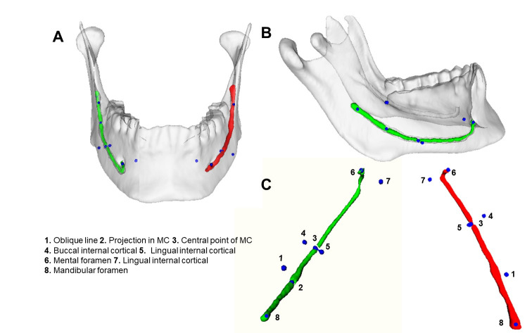
Materials and method: 75 CBCT images were obtained from a secondary database, and 3D analysis was performed using ITK-SNAP and 3D Slicer software. The 3D evaluation consisted of determining the orientation of the position of the mandible, segmentation of the mandible and the MC, creating 3D models, and establishing anatomical landmarks. Vertical (supero-inferior, SI), transverse (mediolateral, RL,) and 3D measurements were performed.
Results: The position of the MC is modified according to the skeletal pattern and by morphological factors of the mandible such as sex and gonial angle. The proximity of the MC to the oblique line is smaller in the SI direction in Class III, and the position of the MC is associated with variation in the gonial angle. It may be closer to the cortical lingual in the central region.
Conclusion: The mandibular canal position should be considered in tomographic evaluation during diagnosis and therapeutic planning of mandible surgeries, especially in cases of sagittal ramus osteotomy.

 求助内容:
求助内容: 应助结果提醒方式:


