{"title":"使用二维和三维计算机断层成像技术测量手工关节盂形状的比较分析。","authors":"Maxwell S Renna, Ashley I Simpson","doi":"10.5397/cise.2025.00318","DOIUrl":null,"url":null,"abstract":"<p><strong>Background: </strong>Accurate measurement of glenoid version is essential for optimal implant positioning in shoulder arthroplasty. This study compared the accuracy and reliability of unformatted two-dimensional computed tomography (2D-CT), formatted 2D-CT, and three-dimensional CT (3D-CT) reconstructions in measuring glenoid version.</p><p><strong>Methods: </strong>Shoulder CT scans for arthroplasty were analyzed retrospectively. Glenoid version was measured at the estimated glenoid midpoint using unformatted and formatted 2D-CT in the scapular plane. Measurements from 3D-CT reconstructions using the Corrected Friedman Method were used as the control. Inter- and intra-observer reliability was calculated as well as minimally detectable difference.</p><p><strong>Results: </strong>Sixty-five CT scans were analyzed (mean age, 61.7 years). Mean glenoid version was -3.48° (standard deviation [SD], 8.7°) on unformatted 2D-CT, -3.27° (SD, 8.15°) on formatted 2D-CT, and -4.25° (SD, 7.92°) on 3D-CT. Although no significant difference in mean values was observed (analysis of variance, P=0.245), formatted 2D-CT measurements were within 6° of 3D-CT in 95.4% of cases versus 83.1% for unformatted 2D-CT (P=0.023). Directional error occurred in 27.7% of unformatted scans and 16.9% of formatted scans. Inter-observer reliability was highest for 3D-CT (intraclass correlation coefficient [ICC]=0.83; 95% CI, 0.74-0.89), and intra-observer agreement was strongest for 3D-CT (ICC=0.91; 95% CI, 0.86-0.94), followed by formatted 2D-CT (ICC=0.83; 95% CI, 0.73-0.89) and unformatted 2D-CT (ICC=0.77; 95% CI, 0.65-0.85).</p><p><strong>Conclusions: </strong>3D-CT reconstructions are widely considered the most accurate and reproducible method for glenoid version assessment, supported by multiple comparative imaging studies. Formatted 2D-CT provides a reliable alternative when 3D-CT is unavailable, significantly outperforming unformatted 2D-CT in both agreement with the 3D reference and intra- and inter-observer reliability. Level of evidence: IV.</p>","PeriodicalId":33981,"journal":{"name":"Clinics in Shoulder and Elbow","volume":" ","pages":""},"PeriodicalIF":1.7000,"publicationDate":"2025-07-31","publicationTypes":"Journal Article","fieldsOfStudy":null,"isOpenAccess":false,"openAccessPdf":"https://www.ncbi.nlm.nih.gov/pmc/articles/PMC12415454/pdf/","citationCount":"0","resultStr":"{\"title\":\"A comparative analysis of manual glenoid version measurement using two-dimensional and three-dimensional computed tomography imaging techniques.\",\"authors\":\"Maxwell S Renna, Ashley I Simpson\",\"doi\":\"10.5397/cise.2025.00318\",\"DOIUrl\":null,\"url\":null,\"abstract\":\"<p><strong>Background: </strong>Accurate measurement of glenoid version is essential for optimal implant positioning in shoulder arthroplasty. This study compared the accuracy and reliability of unformatted two-dimensional computed tomography (2D-CT), formatted 2D-CT, and three-dimensional CT (3D-CT) reconstructions in measuring glenoid version.</p><p><strong>Methods: </strong>Shoulder CT scans for arthroplasty were analyzed retrospectively. Glenoid version was measured at the estimated glenoid midpoint using unformatted and formatted 2D-CT in the scapular plane. Measurements from 3D-CT reconstructions using the Corrected Friedman Method were used as the control. Inter- and intra-observer reliability was calculated as well as minimally detectable difference.</p><p><strong>Results: </strong>Sixty-five CT scans were analyzed (mean age, 61.7 years). Mean glenoid version was -3.48° (standard deviation [SD], 8.7°) on unformatted 2D-CT, -3.27° (SD, 8.15°) on formatted 2D-CT, and -4.25° (SD, 7.92°) on 3D-CT. Although no significant difference in mean values was observed (analysis of variance, P=0.245), formatted 2D-CT measurements were within 6° of 3D-CT in 95.4% of cases versus 83.1% for unformatted 2D-CT (P=0.023). Directional error occurred in 27.7% of unformatted scans and 16.9% of formatted scans. Inter-observer reliability was highest for 3D-CT (intraclass correlation coefficient [ICC]=0.83; 95% CI, 0.74-0.89), and intra-observer agreement was strongest for 3D-CT (ICC=0.91; 95% CI, 0.86-0.94), followed by formatted 2D-CT (ICC=0.83; 95% CI, 0.73-0.89) and unformatted 2D-CT (ICC=0.77; 95% CI, 0.65-0.85).</p><p><strong>Conclusions: </strong>3D-CT reconstructions are widely considered the most accurate and reproducible method for glenoid version assessment, supported by multiple comparative imaging studies. Formatted 2D-CT provides a reliable alternative when 3D-CT is unavailable, significantly outperforming unformatted 2D-CT in both agreement with the 3D reference and intra- and inter-observer reliability. Level of evidence: IV.</p>\",\"PeriodicalId\":33981,\"journal\":{\"name\":\"Clinics in Shoulder and Elbow\",\"volume\":\" \",\"pages\":\"\"},\"PeriodicalIF\":1.7000,\"publicationDate\":\"2025-07-31\",\"publicationTypes\":\"Journal Article\",\"fieldsOfStudy\":null,\"isOpenAccess\":false,\"openAccessPdf\":\"https://www.ncbi.nlm.nih.gov/pmc/articles/PMC12415454/pdf/\",\"citationCount\":\"0\",\"resultStr\":null,\"platform\":\"Semanticscholar\",\"paperid\":null,\"PeriodicalName\":\"Clinics in Shoulder and Elbow\",\"FirstCategoryId\":\"1085\",\"ListUrlMain\":\"https://doi.org/10.5397/cise.2025.00318\",\"RegionNum\":0,\"RegionCategory\":null,\"ArticlePicture\":[],\"TitleCN\":null,\"AbstractTextCN\":null,\"PMCID\":null,\"EPubDate\":\"\",\"PubModel\":\"\",\"JCR\":\"Q2\",\"JCRName\":\"ORTHOPEDICS\",\"Score\":null,\"Total\":0}","platform":"Semanticscholar","paperid":null,"PeriodicalName":"Clinics in Shoulder and Elbow","FirstCategoryId":"1085","ListUrlMain":"https://doi.org/10.5397/cise.2025.00318","RegionNum":0,"RegionCategory":null,"ArticlePicture":[],"TitleCN":null,"AbstractTextCN":null,"PMCID":null,"EPubDate":"","PubModel":"","JCR":"Q2","JCRName":"ORTHOPEDICS","Score":null,"Total":0}
A comparative analysis of manual glenoid version measurement using two-dimensional and three-dimensional computed tomography imaging techniques.
Background: Accurate measurement of glenoid version is essential for optimal implant positioning in shoulder arthroplasty. This study compared the accuracy and reliability of unformatted two-dimensional computed tomography (2D-CT), formatted 2D-CT, and three-dimensional CT (3D-CT) reconstructions in measuring glenoid version.
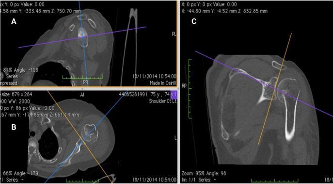
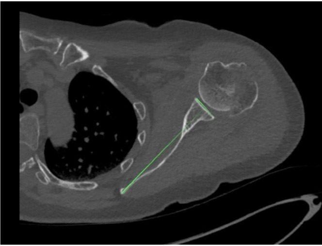
Methods: Shoulder CT scans for arthroplasty were analyzed retrospectively. Glenoid version was measured at the estimated glenoid midpoint using unformatted and formatted 2D-CT in the scapular plane. Measurements from 3D-CT reconstructions using the Corrected Friedman Method were used as the control. Inter- and intra-observer reliability was calculated as well as minimally detectable difference.
Results: Sixty-five CT scans were analyzed (mean age, 61.7 years). Mean glenoid version was -3.48° (standard deviation [SD], 8.7°) on unformatted 2D-CT, -3.27° (SD, 8.15°) on formatted 2D-CT, and -4.25° (SD, 7.92°) on 3D-CT. Although no significant difference in mean values was observed (analysis of variance, P=0.245), formatted 2D-CT measurements were within 6° of 3D-CT in 95.4% of cases versus 83.1% for unformatted 2D-CT (P=0.023). Directional error occurred in 27.7% of unformatted scans and 16.9% of formatted scans. Inter-observer reliability was highest for 3D-CT (intraclass correlation coefficient [ICC]=0.83; 95% CI, 0.74-0.89), and intra-observer agreement was strongest for 3D-CT (ICC=0.91; 95% CI, 0.86-0.94), followed by formatted 2D-CT (ICC=0.83; 95% CI, 0.73-0.89) and unformatted 2D-CT (ICC=0.77; 95% CI, 0.65-0.85).
Conclusions: 3D-CT reconstructions are widely considered the most accurate and reproducible method for glenoid version assessment, supported by multiple comparative imaging studies. Formatted 2D-CT provides a reliable alternative when 3D-CT is unavailable, significantly outperforming unformatted 2D-CT in both agreement with the 3D reference and intra- and inter-observer reliability. Level of evidence: IV.

 求助内容:
求助内容: 应助结果提醒方式:


