Ming Guo, Bingjie Li, Jun Zhao, Chen Bai, Weiyong Yu, Hongxia Zhang, Haoyuan Li, Yongxue Yuan, Qingsu Zhang, Tong Zhang
{"title":"基于静息态fMRI的脑梗死后吞咽困难患者岛叶形态学及功能连通性分析。","authors":"Ming Guo, Bingjie Li, Jun Zhao, Chen Bai, Weiyong Yu, Hongxia Zhang, Haoyuan Li, Yongxue Yuan, Qingsu Zhang, Tong Zhang","doi":"10.1186/s12883-025-04322-1","DOIUrl":null,"url":null,"abstract":"<p><strong>Objective: </strong>The insula, as a critical hub for multimodal information integration, plays a pivotal role in post-stroke dysphagia(PSD). However, the mechanisms underlying its structural and functional network reorganization remain elusive. This study aims to systematically investigate the alterations in gray matter volume and functional connectivity patterns of the insula in patients with dysphagia after cerebral infarction using multimodal neuroimaging techniques, and to untangle their clinical associations with swallowing function impairments.</p><p><strong>Methods: </strong>Three groups of subjects were recruited: healthy controls (HC, n = 15), cerebral infarction patients without dysphagia (ND, n = 13), and cerebral infarction patients with dysphagia (DYS, n = 11). Resting-state functional magnetic resonance imaging (rs-fMRI) and high-resolution T1-weighted structural imaging data were acquired. Seed-based analysis (using the CONN FC toolbox) was employed to quantify the whole-brain functional connectivity (FC) of the insula, and voxel-based morphometry (VBM) was used to assess gray matter volume changes. Swallowing function was standardized using the Fiberoptic Endoscopic Evaluation of Swallowing (FEES) and the Penetration/Aspiration Scale (PAS).</p><p><strong>Results: </strong>The DYS, ND, and HC groups showed significant differences in grey matter volume in the left insula (pFDR =0.041). Compared to the HC group, both cerebral infarction groups (ND and DYS) demonstrated increased functional connectivity between the left insula and the left lateral occipital cortex (superior division), left precuneus, and left cerebellum. In contrast, functional connectivity with the right insula cortex, right frontal operculum cortex, left anterior cingulate, and right frontal pole was decreased. Among these differences, compared to the ND group, the DYS group showed a more significant reduction in functional connectivity within the right frontal operculum cortex and a more pronounced increase in functional connectivity within the left lateral occipital cortex superior division and left cerebellum. Compared to the HC group, patients in both cerebral infarction groups (ND and DYS) showed significantly enhanced functional connectivity between the right insula and the right posterior cingulate gyrus, left lateral occipital cortex (superior division), right precuneus, left frontal pole and right frontal pole. Conversely, functional connectivity with the left insula cortex and left anterior cingulate gyrus was significantly reduced. Moreover, compared to the ND group, the DYS group demonstrated more pronounced increases in functional connectivity within the right posterior cingulate gyrus and right superior cerebellar peduncle, along with a more significant decrease in functional connectivity within the right insula cortex. Enhanced FC between the left insula and the left lateral occipital cortex (superior division) correlated positively with PAS, while enhanced FC between the right insula and the right cerebellum correlated negatively with PAS.</p><p><strong>Conclusion: </strong>Our study found left insular gray matter atrophy underlies the pathology of PSD, and abnormal insular functional connectivity is key to its development. The severity of post-stroke dysphagia can affect the functional connectivity between the insula and the right cerebellum as well as the left occipital lobe. These results reveal potential neural compensation mechanisms in PSD and offer new directions for clinical prognostic biomarker development.</p>","PeriodicalId":9170,"journal":{"name":"BMC Neurology","volume":"25 1","pages":"307"},"PeriodicalIF":2.2000,"publicationDate":"2025-07-30","publicationTypes":"Journal Article","fieldsOfStudy":null,"isOpenAccess":false,"openAccessPdf":"https://www.ncbi.nlm.nih.gov/pmc/articles/PMC12309065/pdf/","citationCount":"0","resultStr":"{\"title\":\"Morphological analysis and functional connectivity of the insular in patients with dysphagia after cerebral infarction based on resting-state fMRI.\",\"authors\":\"Ming Guo, Bingjie Li, Jun Zhao, Chen Bai, Weiyong Yu, Hongxia Zhang, Haoyuan Li, Yongxue Yuan, Qingsu Zhang, Tong Zhang\",\"doi\":\"10.1186/s12883-025-04322-1\",\"DOIUrl\":null,\"url\":null,\"abstract\":\"<p><strong>Objective: </strong>The insula, as a critical hub for multimodal information integration, plays a pivotal role in post-stroke dysphagia(PSD). However, the mechanisms underlying its structural and functional network reorganization remain elusive. This study aims to systematically investigate the alterations in gray matter volume and functional connectivity patterns of the insula in patients with dysphagia after cerebral infarction using multimodal neuroimaging techniques, and to untangle their clinical associations with swallowing function impairments.</p><p><strong>Methods: </strong>Three groups of subjects were recruited: healthy controls (HC, n = 15), cerebral infarction patients without dysphagia (ND, n = 13), and cerebral infarction patients with dysphagia (DYS, n = 11). Resting-state functional magnetic resonance imaging (rs-fMRI) and high-resolution T1-weighted structural imaging data were acquired. Seed-based analysis (using the CONN FC toolbox) was employed to quantify the whole-brain functional connectivity (FC) of the insula, and voxel-based morphometry (VBM) was used to assess gray matter volume changes. Swallowing function was standardized using the Fiberoptic Endoscopic Evaluation of Swallowing (FEES) and the Penetration/Aspiration Scale (PAS).</p><p><strong>Results: </strong>The DYS, ND, and HC groups showed significant differences in grey matter volume in the left insula (pFDR =0.041). Compared to the HC group, both cerebral infarction groups (ND and DYS) demonstrated increased functional connectivity between the left insula and the left lateral occipital cortex (superior division), left precuneus, and left cerebellum. In contrast, functional connectivity with the right insula cortex, right frontal operculum cortex, left anterior cingulate, and right frontal pole was decreased. Among these differences, compared to the ND group, the DYS group showed a more significant reduction in functional connectivity within the right frontal operculum cortex and a more pronounced increase in functional connectivity within the left lateral occipital cortex superior division and left cerebellum. Compared to the HC group, patients in both cerebral infarction groups (ND and DYS) showed significantly enhanced functional connectivity between the right insula and the right posterior cingulate gyrus, left lateral occipital cortex (superior division), right precuneus, left frontal pole and right frontal pole. Conversely, functional connectivity with the left insula cortex and left anterior cingulate gyrus was significantly reduced. Moreover, compared to the ND group, the DYS group demonstrated more pronounced increases in functional connectivity within the right posterior cingulate gyrus and right superior cerebellar peduncle, along with a more significant decrease in functional connectivity within the right insula cortex. Enhanced FC between the left insula and the left lateral occipital cortex (superior division) correlated positively with PAS, while enhanced FC between the right insula and the right cerebellum correlated negatively with PAS.</p><p><strong>Conclusion: </strong>Our study found left insular gray matter atrophy underlies the pathology of PSD, and abnormal insular functional connectivity is key to its development. The severity of post-stroke dysphagia can affect the functional connectivity between the insula and the right cerebellum as well as the left occipital lobe. These results reveal potential neural compensation mechanisms in PSD and offer new directions for clinical prognostic biomarker development.</p>\",\"PeriodicalId\":9170,\"journal\":{\"name\":\"BMC Neurology\",\"volume\":\"25 1\",\"pages\":\"307\"},\"PeriodicalIF\":2.2000,\"publicationDate\":\"2025-07-30\",\"publicationTypes\":\"Journal Article\",\"fieldsOfStudy\":null,\"isOpenAccess\":false,\"openAccessPdf\":\"https://www.ncbi.nlm.nih.gov/pmc/articles/PMC12309065/pdf/\",\"citationCount\":\"0\",\"resultStr\":null,\"platform\":\"Semanticscholar\",\"paperid\":null,\"PeriodicalName\":\"BMC Neurology\",\"FirstCategoryId\":\"3\",\"ListUrlMain\":\"https://doi.org/10.1186/s12883-025-04322-1\",\"RegionNum\":3,\"RegionCategory\":\"医学\",\"ArticlePicture\":[],\"TitleCN\":null,\"AbstractTextCN\":null,\"PMCID\":null,\"EPubDate\":\"\",\"PubModel\":\"\",\"JCR\":\"Q3\",\"JCRName\":\"CLINICAL NEUROLOGY\",\"Score\":null,\"Total\":0}","platform":"Semanticscholar","paperid":null,"PeriodicalName":"BMC Neurology","FirstCategoryId":"3","ListUrlMain":"https://doi.org/10.1186/s12883-025-04322-1","RegionNum":3,"RegionCategory":"医学","ArticlePicture":[],"TitleCN":null,"AbstractTextCN":null,"PMCID":null,"EPubDate":"","PubModel":"","JCR":"Q3","JCRName":"CLINICAL NEUROLOGY","Score":null,"Total":0}
引用次数: 0
摘要
目的:脑岛作为脑卒中后吞咽困难(PSD)的多模态信息整合关键枢纽,在脑卒中后吞咽困难(PSD)中起着关键作用。然而,其结构和功能网络重组的机制尚不清楚。本研究旨在利用多模态神经影像学技术系统研究脑梗死后吞咽困难患者脑岛灰质体积和功能连接模式的改变,并阐明其与吞咽功能障碍的临床关联。方法:选取健康对照组(HC, n = 15)、无吞咽困难的脑梗死患者(ND, n = 13)和有吞咽困难的脑梗死患者(DYS, n = 11)为研究对象。静息状态功能磁共振成像(rs-fMRI)和高分辨率t1加权结构成像数据。采用基于种子的分析(使用CONN FC工具箱)来量化脑岛的全脑功能连接(FC),采用基于体素的形态测量(VBM)来评估灰质体积变化。使用光纤内镜吞咽评估(FEES)和穿透/吸入量表(PAS)对吞咽功能进行标准化。结果:DYS、ND、HC组左岛灰质体积差异有统计学意义(pFDR =0.041)。与HC组相比,两个脑梗死组(ND和DYS)均表现出左侧脑岛与左侧枕外侧皮质(上分)、左侧楔前叶和左侧小脑之间的功能连通性增加。与此相反,与右岛叶皮层、右额盖皮层、左前扣带和右额极的功能连通性下降。在这些差异中,与ND组相比,DYS组显示右侧额盖皮层内功能连通性更显着降低,左侧外侧枕皮质上分区和左侧小脑内功能连通性更显着增加。与HC组相比,两组脑梗死患者(ND组和DYS组)的右侧脑岛与右侧扣带回后回、左侧枕外侧皮层(上分)、右侧楔前叶、左额极和右额极之间的功能连连性均显著增强。相反,与左脑岛皮层和左前扣带回的功能连通性明显降低。此外,与ND组相比,DYS组右侧扣带后回和右侧小脑上蒂的功能连通性增加更为明显,而右侧脑岛皮层的功能连通性下降更为明显。左侧脑岛与左侧枕外侧皮质(上分)之间的FC增强与PAS呈正相关,而右侧脑岛与右侧小脑之间的FC增强与PAS负相关。结论:我们的研究发现左岛灰质萎缩是PSD的病理基础,而异常的岛功能连接是其发展的关键。脑卒中后吞咽困难的严重程度可影响脑岛与右小脑以及左枕叶之间的功能连接。这些结果揭示了PSD中潜在的神经代偿机制,并为临床预后生物标志物的开发提供了新的方向。
Morphological analysis and functional connectivity of the insular in patients with dysphagia after cerebral infarction based on resting-state fMRI.
Objective: The insula, as a critical hub for multimodal information integration, plays a pivotal role in post-stroke dysphagia(PSD). However, the mechanisms underlying its structural and functional network reorganization remain elusive. This study aims to systematically investigate the alterations in gray matter volume and functional connectivity patterns of the insula in patients with dysphagia after cerebral infarction using multimodal neuroimaging techniques, and to untangle their clinical associations with swallowing function impairments.
Methods: Three groups of subjects were recruited: healthy controls (HC, n = 15), cerebral infarction patients without dysphagia (ND, n = 13), and cerebral infarction patients with dysphagia (DYS, n = 11). Resting-state functional magnetic resonance imaging (rs-fMRI) and high-resolution T1-weighted structural imaging data were acquired. Seed-based analysis (using the CONN FC toolbox) was employed to quantify the whole-brain functional connectivity (FC) of the insula, and voxel-based morphometry (VBM) was used to assess gray matter volume changes. Swallowing function was standardized using the Fiberoptic Endoscopic Evaluation of Swallowing (FEES) and the Penetration/Aspiration Scale (PAS).
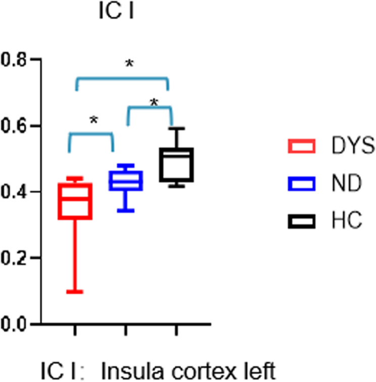
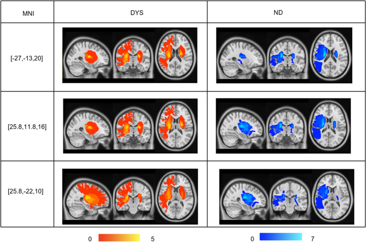
Results: The DYS, ND, and HC groups showed significant differences in grey matter volume in the left insula (pFDR =0.041). Compared to the HC group, both cerebral infarction groups (ND and DYS) demonstrated increased functional connectivity between the left insula and the left lateral occipital cortex (superior division), left precuneus, and left cerebellum. In contrast, functional connectivity with the right insula cortex, right frontal operculum cortex, left anterior cingulate, and right frontal pole was decreased. Among these differences, compared to the ND group, the DYS group showed a more significant reduction in functional connectivity within the right frontal operculum cortex and a more pronounced increase in functional connectivity within the left lateral occipital cortex superior division and left cerebellum. Compared to the HC group, patients in both cerebral infarction groups (ND and DYS) showed significantly enhanced functional connectivity between the right insula and the right posterior cingulate gyrus, left lateral occipital cortex (superior division), right precuneus, left frontal pole and right frontal pole. Conversely, functional connectivity with the left insula cortex and left anterior cingulate gyrus was significantly reduced. Moreover, compared to the ND group, the DYS group demonstrated more pronounced increases in functional connectivity within the right posterior cingulate gyrus and right superior cerebellar peduncle, along with a more significant decrease in functional connectivity within the right insula cortex. Enhanced FC between the left insula and the left lateral occipital cortex (superior division) correlated positively with PAS, while enhanced FC between the right insula and the right cerebellum correlated negatively with PAS.
Conclusion: Our study found left insular gray matter atrophy underlies the pathology of PSD, and abnormal insular functional connectivity is key to its development. The severity of post-stroke dysphagia can affect the functional connectivity between the insula and the right cerebellum as well as the left occipital lobe. These results reveal potential neural compensation mechanisms in PSD and offer new directions for clinical prognostic biomarker development.
期刊介绍:
BMC Neurology is an open access, peer-reviewed journal that considers articles on all aspects of the prevention, diagnosis and management of neurological disorders, as well as related molecular genetics, pathophysiology, and epidemiology.

 求助内容:
求助内容: 应助结果提醒方式:


