AmirHossein RahimBakhsh, Asma Kheirollahi, Akram Vatannejad, Sara Shokrpoor, Rahman Mohammadi
{"title":"s -腺苷甲硫氨酸对stz诱导的糖尿病大鼠氧化应激及组织损伤的保护作用。","authors":"AmirHossein RahimBakhsh, Asma Kheirollahi, Akram Vatannejad, Sara Shokrpoor, Rahman Mohammadi","doi":"10.1007/s00726-025-03471-4","DOIUrl":null,"url":null,"abstract":"<div><h3>Background</h3><p>Oxidative stress is a key contributor to the progression of diabetes mellitus and its associated complications. Recently, S-adenosyl methionine (SAM) has shown promise in mitigating oxidative stress and improving glucose metabolism. This study aimed to investigate the effects of SAM supplementation on biochemical parameters, oxidative stress markers, and histopathological alterations in the kidneys and liver of streptozotocin (STZ)-induced diabetic rats.</p><h3>Methods</h3><p>Eighteen male Wistar rats were randomly divided into three groups (n = 6 per group): non-diabetic control, diabetic control, and diabetic rats treated with SAM (10 mg/kg/day, intraperitoneally) for 4 weeks. Fasting blood glucose, renal and hepatic biochemical markers (urea, creatinine, ALT, AST), and oxidative stress markers (malondialdehyde, protein carbonyls, total antioxidant capacity) were measured. Histopathological changes in kidney and liver tissues were also assessed.</p><h3>Results</h3><p>Diabetic rats treated with SAM exhibited minor, non-significant changes in fasting blood glucose, urea, creatinine, ALT, and AST levels. In contrast, treatment with SAM in diabetic rats significantly reduced malondialdehyde and protein carbonyl levels in both kidney and liver tissues compared to the diabetic control group (P < 0.05). Furthermore, histopathological analysis revealed improved tissue architecture and reduced pathological changes in the diabetic group treated with SAM.</p><h3>Conclusion</h3><p>Our findings demonstrated that SAM supplementation exerts significant antioxidant and histopathological protective effects against diabetes-induced damage in kidney and liver tissues.</p></div>","PeriodicalId":7810,"journal":{"name":"Amino Acids","volume":"57 1","pages":""},"PeriodicalIF":2.4000,"publicationDate":"2025-07-31","publicationTypes":"Journal Article","fieldsOfStudy":null,"isOpenAccess":false,"openAccessPdf":"https://www.ncbi.nlm.nih.gov/pmc/articles/PMC12313745/pdf/","citationCount":"0","resultStr":"{\"title\":\"Protective effects of S-adenosyl methionine on oxidative stress and tissue damage in STZ-induced diabetic rats\",\"authors\":\"AmirHossein RahimBakhsh, Asma Kheirollahi, Akram Vatannejad, Sara Shokrpoor, Rahman Mohammadi\",\"doi\":\"10.1007/s00726-025-03471-4\",\"DOIUrl\":null,\"url\":null,\"abstract\":\"<div><h3>Background</h3><p>Oxidative stress is a key contributor to the progression of diabetes mellitus and its associated complications. Recently, S-adenosyl methionine (SAM) has shown promise in mitigating oxidative stress and improving glucose metabolism. This study aimed to investigate the effects of SAM supplementation on biochemical parameters, oxidative stress markers, and histopathological alterations in the kidneys and liver of streptozotocin (STZ)-induced diabetic rats.</p><h3>Methods</h3><p>Eighteen male Wistar rats were randomly divided into three groups (n = 6 per group): non-diabetic control, diabetic control, and diabetic rats treated with SAM (10 mg/kg/day, intraperitoneally) for 4 weeks. Fasting blood glucose, renal and hepatic biochemical markers (urea, creatinine, ALT, AST), and oxidative stress markers (malondialdehyde, protein carbonyls, total antioxidant capacity) were measured. Histopathological changes in kidney and liver tissues were also assessed.</p><h3>Results</h3><p>Diabetic rats treated with SAM exhibited minor, non-significant changes in fasting blood glucose, urea, creatinine, ALT, and AST levels. In contrast, treatment with SAM in diabetic rats significantly reduced malondialdehyde and protein carbonyl levels in both kidney and liver tissues compared to the diabetic control group (P < 0.05). Furthermore, histopathological analysis revealed improved tissue architecture and reduced pathological changes in the diabetic group treated with SAM.</p><h3>Conclusion</h3><p>Our findings demonstrated that SAM supplementation exerts significant antioxidant and histopathological protective effects against diabetes-induced damage in kidney and liver tissues.</p></div>\",\"PeriodicalId\":7810,\"journal\":{\"name\":\"Amino Acids\",\"volume\":\"57 1\",\"pages\":\"\"},\"PeriodicalIF\":2.4000,\"publicationDate\":\"2025-07-31\",\"publicationTypes\":\"Journal Article\",\"fieldsOfStudy\":null,\"isOpenAccess\":false,\"openAccessPdf\":\"https://www.ncbi.nlm.nih.gov/pmc/articles/PMC12313745/pdf/\",\"citationCount\":\"0\",\"resultStr\":null,\"platform\":\"Semanticscholar\",\"paperid\":null,\"PeriodicalName\":\"Amino Acids\",\"FirstCategoryId\":\"99\",\"ListUrlMain\":\"https://link.springer.com/article/10.1007/s00726-025-03471-4\",\"RegionNum\":3,\"RegionCategory\":\"生物学\",\"ArticlePicture\":[],\"TitleCN\":null,\"AbstractTextCN\":null,\"PMCID\":null,\"EPubDate\":\"\",\"PubModel\":\"\",\"JCR\":\"Q3\",\"JCRName\":\"BIOCHEMISTRY & MOLECULAR BIOLOGY\",\"Score\":null,\"Total\":0}","platform":"Semanticscholar","paperid":null,"PeriodicalName":"Amino Acids","FirstCategoryId":"99","ListUrlMain":"https://link.springer.com/article/10.1007/s00726-025-03471-4","RegionNum":3,"RegionCategory":"生物学","ArticlePicture":[],"TitleCN":null,"AbstractTextCN":null,"PMCID":null,"EPubDate":"","PubModel":"","JCR":"Q3","JCRName":"BIOCHEMISTRY & MOLECULAR BIOLOGY","Score":null,"Total":0}
Protective effects of S-adenosyl methionine on oxidative stress and tissue damage in STZ-induced diabetic rats
Background
Oxidative stress is a key contributor to the progression of diabetes mellitus and its associated complications. Recently, S-adenosyl methionine (SAM) has shown promise in mitigating oxidative stress and improving glucose metabolism. This study aimed to investigate the effects of SAM supplementation on biochemical parameters, oxidative stress markers, and histopathological alterations in the kidneys and liver of streptozotocin (STZ)-induced diabetic rats.
Methods
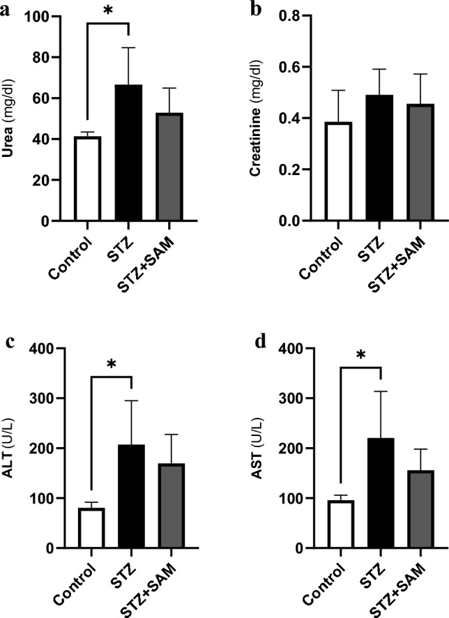
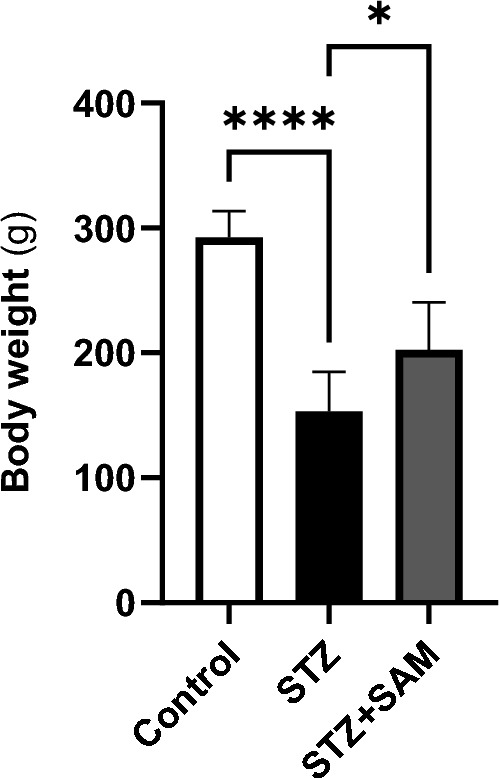
Eighteen male Wistar rats were randomly divided into three groups (n = 6 per group): non-diabetic control, diabetic control, and diabetic rats treated with SAM (10 mg/kg/day, intraperitoneally) for 4 weeks. Fasting blood glucose, renal and hepatic biochemical markers (urea, creatinine, ALT, AST), and oxidative stress markers (malondialdehyde, protein carbonyls, total antioxidant capacity) were measured. Histopathological changes in kidney and liver tissues were also assessed.
Results
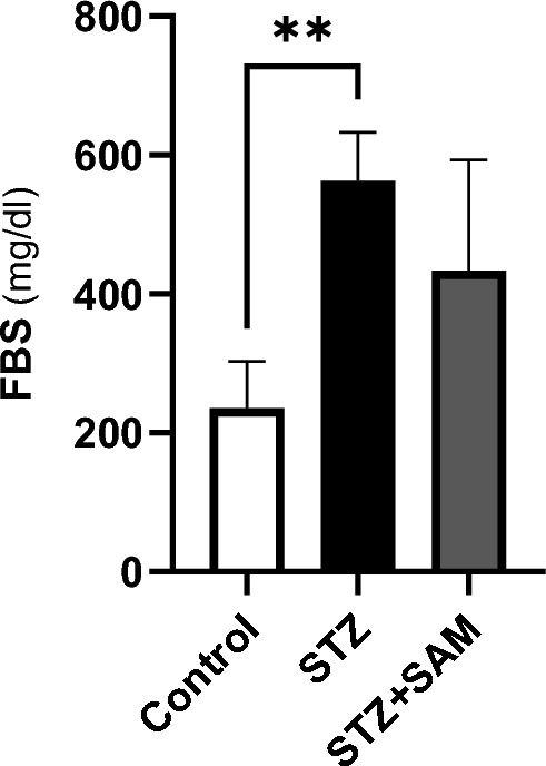
Diabetic rats treated with SAM exhibited minor, non-significant changes in fasting blood glucose, urea, creatinine, ALT, and AST levels. In contrast, treatment with SAM in diabetic rats significantly reduced malondialdehyde and protein carbonyl levels in both kidney and liver tissues compared to the diabetic control group (P < 0.05). Furthermore, histopathological analysis revealed improved tissue architecture and reduced pathological changes in the diabetic group treated with SAM.
Conclusion
Our findings demonstrated that SAM supplementation exerts significant antioxidant and histopathological protective effects against diabetes-induced damage in kidney and liver tissues.
期刊介绍:
Amino Acids publishes contributions from all fields of amino acid and protein research: analysis, separation, synthesis, biosynthesis, cross linking amino acids, racemization/enantiomers, modification of amino acids as phosphorylation, methylation, acetylation, glycosylation and nonenzymatic glycosylation, new roles for amino acids in physiology and pathophysiology, biology, amino acid analogues and derivatives, polyamines, radiated amino acids, peptides, stable isotopes and isotopes of amino acids. Applications in medicine, food chemistry, nutrition, gastroenterology, nephrology, neurochemistry, pharmacology, excitatory amino acids are just some of the topics covered. Fields of interest include: Biochemistry, food chemistry, nutrition, neurology, psychiatry, pharmacology, nephrology, gastroenterology, microbiology
 求助内容:
求助内容: 应助结果提醒方式:


