Meiqi Weng , Chenxi Zhang , Xunbo Miao , Chuangzheng Yang , Mei Cao , Nan Sun , Suxing Liu , Xingqiang Wang , Jinyong Zhang
{"title":"中国细棘棘球绦虫(甲壳纲:棘球绦虫科)下表皮的小孢子虫","authors":"Meiqi Weng , Chenxi Zhang , Xunbo Miao , Chuangzheng Yang , Mei Cao , Nan Sun , Suxing Liu , Xingqiang Wang , Jinyong Zhang","doi":"10.1016/j.jip.2025.108414","DOIUrl":null,"url":null,"abstract":"<div><div>A new microsporidian species was described from the hypoderm of <em>Diaphanosoma leuchtenbergianum</em> sampled from a eutrophic water body of Changshu city, Jiangsu province, China. Infected daphnia generally appeared opaque due to numerous spores accumulated in the host integument. The earliest developmental stages observed were uninucleate meronts which were in direct contact with the host cell cytoplasm. Multinucleate sporogonial plasmodia developed into uninucleate sporoblasts by plasmotomy. Mature spores were ovoid and monokaryotic, measuring 4.65 ± 0.21 (4.28–5.24) µm long and 2.49 ± 0.15 (2.12–2.82) µm wide. Isofilar polar filament coiled 8–11 turns and arranged in 2–3 rows. Bipartite polaroplast consisted of the tightly packed anterior lamellae and posterior tubules. The exospore was covered with tubular structures and was composed of two layers. Phylogenetic analysis based on the obtained SSU rDNA sequence indicated that the present species clustered with three <em>Neoflabelliforma</em> species (<em>N. dubium</em>, <em>N. aurantiae</em>, and <em>N. leuchtenbergianum</em>) and an unidentified microsporidium from soil with high support values to form an independent branch within canonical Microsporidia. Based on the morphological characters and ultrastructural features, as well as SSU rDNA-inferred phylogenetic relationships, a new species was erected and named as <em>Neoflabelliforma diaphanosoma</em> n. sp.</div></div>","PeriodicalId":16296,"journal":{"name":"Journal of invertebrate pathology","volume":"213 ","pages":"Article 108414"},"PeriodicalIF":2.4000,"publicationDate":"2025-07-30","publicationTypes":"Journal Article","fieldsOfStudy":null,"isOpenAccess":false,"openAccessPdf":"","citationCount":"0","resultStr":"{\"title\":\"Neoflabelliforma diaphanosoma n. sp. (Microsporidia) from hypoderm of diaphanosoma leuchtenbergianum (Crustacea: Sididae) in China\",\"authors\":\"Meiqi Weng , Chenxi Zhang , Xunbo Miao , Chuangzheng Yang , Mei Cao , Nan Sun , Suxing Liu , Xingqiang Wang , Jinyong Zhang\",\"doi\":\"10.1016/j.jip.2025.108414\",\"DOIUrl\":null,\"url\":null,\"abstract\":\"<div><div>A new microsporidian species was described from the hypoderm of <em>Diaphanosoma leuchtenbergianum</em> sampled from a eutrophic water body of Changshu city, Jiangsu province, China. Infected daphnia generally appeared opaque due to numerous spores accumulated in the host integument. The earliest developmental stages observed were uninucleate meronts which were in direct contact with the host cell cytoplasm. Multinucleate sporogonial plasmodia developed into uninucleate sporoblasts by plasmotomy. Mature spores were ovoid and monokaryotic, measuring 4.65 ± 0.21 (4.28–5.24) µm long and 2.49 ± 0.15 (2.12–2.82) µm wide. Isofilar polar filament coiled 8–11 turns and arranged in 2–3 rows. Bipartite polaroplast consisted of the tightly packed anterior lamellae and posterior tubules. The exospore was covered with tubular structures and was composed of two layers. Phylogenetic analysis based on the obtained SSU rDNA sequence indicated that the present species clustered with three <em>Neoflabelliforma</em> species (<em>N. dubium</em>, <em>N. aurantiae</em>, and <em>N. leuchtenbergianum</em>) and an unidentified microsporidium from soil with high support values to form an independent branch within canonical Microsporidia. Based on the morphological characters and ultrastructural features, as well as SSU rDNA-inferred phylogenetic relationships, a new species was erected and named as <em>Neoflabelliforma diaphanosoma</em> n. sp.</div></div>\",\"PeriodicalId\":16296,\"journal\":{\"name\":\"Journal of invertebrate pathology\",\"volume\":\"213 \",\"pages\":\"Article 108414\"},\"PeriodicalIF\":2.4000,\"publicationDate\":\"2025-07-30\",\"publicationTypes\":\"Journal Article\",\"fieldsOfStudy\":null,\"isOpenAccess\":false,\"openAccessPdf\":\"\",\"citationCount\":\"0\",\"resultStr\":null,\"platform\":\"Semanticscholar\",\"paperid\":null,\"PeriodicalName\":\"Journal of invertebrate pathology\",\"FirstCategoryId\":\"99\",\"ListUrlMain\":\"https://www.sciencedirect.com/science/article/pii/S002220112500148X\",\"RegionNum\":3,\"RegionCategory\":\"生物学\",\"ArticlePicture\":[],\"TitleCN\":null,\"AbstractTextCN\":null,\"PMCID\":null,\"EPubDate\":\"\",\"PubModel\":\"\",\"JCR\":\"Q1\",\"JCRName\":\"ZOOLOGY\",\"Score\":null,\"Total\":0}","platform":"Semanticscholar","paperid":null,"PeriodicalName":"Journal of invertebrate pathology","FirstCategoryId":"99","ListUrlMain":"https://www.sciencedirect.com/science/article/pii/S002220112500148X","RegionNum":3,"RegionCategory":"生物学","ArticlePicture":[],"TitleCN":null,"AbstractTextCN":null,"PMCID":null,"EPubDate":"","PubModel":"","JCR":"Q1","JCRName":"ZOOLOGY","Score":null,"Total":0}
引用次数: 0
摘要
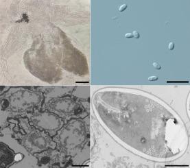
在江苏省常熟市富营养化水体中,发现了一种微孢子虫新种。受感染的水蚤通常表现为不透明,因为大量孢子积聚在寄主被皮里。最早观察到的发育阶段是与宿主细胞质直接接触的无核小体。多核孢子胞经胞浆切开术发育成单核孢子母细胞。成熟孢子长4.65±0.21(4.28-5.24)µm,宽2.49±0.15(2.12-2.82)µm,为卵球形单核细胞。等丝极性灯丝盘绕8-11圈,排列成2-3行。两节极质体由紧密排列的前片和后小管组成。外孢子被管状结构覆盖,由两层结构组成。基于获得的SSU rDNA序列的系统发育分析表明,本种与3个新flabelliforma种(N. dubium、N. aurantiae和N. leuchtenbergianum)和一个来自土壤的未识别的高支持值微孢子虫聚集在一起,在标准微孢子虫中形成一个独立的分支。根据其形态特征和超微结构特征,以及SSU rdna推断的系统发育关系,建立了一个新种,命名为Neoflabelliforma diaphanosoma n. sp。
Neoflabelliforma diaphanosoma n. sp. (Microsporidia) from hypoderm of diaphanosoma leuchtenbergianum (Crustacea: Sididae) in China
A new microsporidian species was described from the hypoderm of Diaphanosoma leuchtenbergianum sampled from a eutrophic water body of Changshu city, Jiangsu province, China. Infected daphnia generally appeared opaque due to numerous spores accumulated in the host integument. The earliest developmental stages observed were uninucleate meronts which were in direct contact with the host cell cytoplasm. Multinucleate sporogonial plasmodia developed into uninucleate sporoblasts by plasmotomy. Mature spores were ovoid and monokaryotic, measuring 4.65 ± 0.21 (4.28–5.24) µm long and 2.49 ± 0.15 (2.12–2.82) µm wide. Isofilar polar filament coiled 8–11 turns and arranged in 2–3 rows. Bipartite polaroplast consisted of the tightly packed anterior lamellae and posterior tubules. The exospore was covered with tubular structures and was composed of two layers. Phylogenetic analysis based on the obtained SSU rDNA sequence indicated that the present species clustered with three Neoflabelliforma species (N. dubium, N. aurantiae, and N. leuchtenbergianum) and an unidentified microsporidium from soil with high support values to form an independent branch within canonical Microsporidia. Based on the morphological characters and ultrastructural features, as well as SSU rDNA-inferred phylogenetic relationships, a new species was erected and named as Neoflabelliforma diaphanosoma n. sp.
期刊介绍:
The Journal of Invertebrate Pathology presents original research articles and notes on the induction and pathogenesis of diseases of invertebrates, including the suppression of diseases in beneficial species, and the use of diseases in controlling undesirable species. In addition, the journal publishes the results of physiological, morphological, genetic, immunological and ecological studies as related to the etiologic agents of diseases of invertebrates.
The Journal of Invertebrate Pathology is the adopted journal of the Society for Invertebrate Pathology, and is available to SIP members at a special reduced price.