L.A. Lefol , A. Sodano , P. Bawuah , J.A. Zeitler , J. Verin , F. Danede , J.F. Willart , J. Siepmann , F. Siepmann
{"title":"
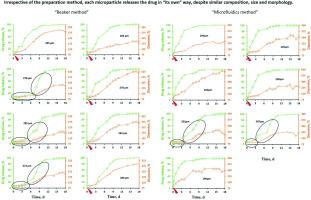
使用微流体装置或烧杯制备的PLGA微粒的释放机制","authors":"L.A. Lefol , A. Sodano , P. Bawuah , J.A. Zeitler , J. Verin , F. Danede , J.F. Willart , J. Siepmann , F. Siepmann","doi":"10.1016/j.ijpx.2025.100366","DOIUrl":null,"url":null,"abstract":"<div><div>The aim of this study was to better understand the release mechanisms of poly(lactic-<em>co</em>-glycolic acid) (PLGA) microparticles prepared via emulsification - solvent extraction/evaporation using a “classical beaker” vs. a “microfluidics device”. Ibuprofen-loaded microparticles were studied by optical microscopy, SEM, X-ray powder diffraction, X-ray μCT and drug release measurements from <em>single</em> microparticles in well agitated phosphate buffer pH 7.4 or agarose gel (mimicking living tissue). The use of a microfluidics device facilitated the preparation of microparticles with a less broad size distribution. However, in addition to the microparticle size, the inner system structure was found to be also of utmost importance for the resulting drug release kinetics in this case. Interestingly, even microparticles with <em>similar size, composition and inner & outer structure</em> exhibited a <em>broad spectrum of individual drug release patterns</em>. This was true, irrespective of the type of preparation method and experimental release set-up, and could be explained as follows: The investigated microparticles were characterized by a continuous inner pore network and an initially smooth & non-porous surface. Drug release set on as soon as: (i) the pore network got direct access to the release medium (e.g., due to a “weak point” in the PLGA surface layer), or (ii) substantial system swelling started (after a lag-time of several days). Importantly, <em>each</em> microparticle had its own, specific structure, which determined “<em>its way</em>” to release the drug. Furthermore, the experimental conditions were found to be of key importance: The presence of a surrounding agarose gel protected the microparticles from damage caused by convective fluid flow, and hindered microparticle swelling, thus, slowing down drug release.</div></div>","PeriodicalId":14280,"journal":{"name":"International Journal of Pharmaceutics: X","volume":"10 ","pages":"Article 100366"},"PeriodicalIF":6.4000,"publicationDate":"2025-07-23","publicationTypes":"Journal Article","fieldsOfStudy":null,"isOpenAccess":false,"openAccessPdf":"","citationCount":"0","resultStr":"{\"title\":\"Release mechanisms of PLGA microparticles prepared using a microfluidics device or a beaker\",\"authors\":\"L.A. Lefol , A. Sodano , P. Bawuah , J.A. Zeitler , J. Verin , F. Danede , J.F. Willart , J. Siepmann , F. Siepmann\",\"doi\":\"10.1016/j.ijpx.2025.100366\",\"DOIUrl\":null,\"url\":null,\"abstract\":\"<div><div>The aim of this study was to better understand the release mechanisms of poly(lactic-<em>co</em>-glycolic acid) (PLGA) microparticles prepared via emulsification - solvent extraction/evaporation using a “classical beaker” vs. a “microfluidics device”. Ibuprofen-loaded microparticles were studied by optical microscopy, SEM, X-ray powder diffraction, X-ray μCT and drug release measurements from <em>single</em> microparticles in well agitated phosphate buffer pH 7.4 or agarose gel (mimicking living tissue). The use of a microfluidics device facilitated the preparation of microparticles with a less broad size distribution. However, in addition to the microparticle size, the inner system structure was found to be also of utmost importance for the resulting drug release kinetics in this case. Interestingly, even microparticles with <em>similar size, composition and inner & outer structure</em> exhibited a <em>broad spectrum of individual drug release patterns</em>. This was true, irrespective of the type of preparation method and experimental release set-up, and could be explained as follows: The investigated microparticles were characterized by a continuous inner pore network and an initially smooth & non-porous surface. Drug release set on as soon as: (i) the pore network got direct access to the release medium (e.g., due to a “weak point” in the PLGA surface layer), or (ii) substantial system swelling started (after a lag-time of several days). Importantly, <em>each</em> microparticle had its own, specific structure, which determined “<em>its way</em>” to release the drug. Furthermore, the experimental conditions were found to be of key importance: The presence of a surrounding agarose gel protected the microparticles from damage caused by convective fluid flow, and hindered microparticle swelling, thus, slowing down drug release.</div></div>\",\"PeriodicalId\":14280,\"journal\":{\"name\":\"International Journal of Pharmaceutics: X\",\"volume\":\"10 \",\"pages\":\"Article 100366\"},\"PeriodicalIF\":6.4000,\"publicationDate\":\"2025-07-23\",\"publicationTypes\":\"Journal Article\",\"fieldsOfStudy\":null,\"isOpenAccess\":false,\"openAccessPdf\":\"\",\"citationCount\":\"0\",\"resultStr\":null,\"platform\":\"Semanticscholar\",\"paperid\":null,\"PeriodicalName\":\"International Journal of Pharmaceutics: X\",\"FirstCategoryId\":\"3\",\"ListUrlMain\":\"https://www.sciencedirect.com/science/article/pii/S2590156725000519\",\"RegionNum\":2,\"RegionCategory\":\"医学\",\"ArticlePicture\":[],\"TitleCN\":null,\"AbstractTextCN\":null,\"PMCID\":null,\"EPubDate\":\"\",\"PubModel\":\"\",\"JCR\":\"Q1\",\"JCRName\":\"PHARMACOLOGY & PHARMACY\",\"Score\":null,\"Total\":0}","platform":"Semanticscholar","paperid":null,"PeriodicalName":"International Journal of Pharmaceutics: X","FirstCategoryId":"3","ListUrlMain":"https://www.sciencedirect.com/science/article/pii/S2590156725000519","RegionNum":2,"RegionCategory":"医学","ArticlePicture":[],"TitleCN":null,"AbstractTextCN":null,"PMCID":null,"EPubDate":"","PubModel":"","JCR":"Q1","JCRName":"PHARMACOLOGY & PHARMACY","Score":null,"Total":0}
Release mechanisms of PLGA microparticles prepared using a microfluidics device or a beaker
The aim of this study was to better understand the release mechanisms of poly(lactic-co-glycolic acid) (PLGA) microparticles prepared via emulsification - solvent extraction/evaporation using a “classical beaker” vs. a “microfluidics device”. Ibuprofen-loaded microparticles were studied by optical microscopy, SEM, X-ray powder diffraction, X-ray μCT and drug release measurements from single microparticles in well agitated phosphate buffer pH 7.4 or agarose gel (mimicking living tissue). The use of a microfluidics device facilitated the preparation of microparticles with a less broad size distribution. However, in addition to the microparticle size, the inner system structure was found to be also of utmost importance for the resulting drug release kinetics in this case. Interestingly, even microparticles with similar size, composition and inner & outer structure exhibited a broad spectrum of individual drug release patterns. This was true, irrespective of the type of preparation method and experimental release set-up, and could be explained as follows: The investigated microparticles were characterized by a continuous inner pore network and an initially smooth & non-porous surface. Drug release set on as soon as: (i) the pore network got direct access to the release medium (e.g., due to a “weak point” in the PLGA surface layer), or (ii) substantial system swelling started (after a lag-time of several days). Importantly, each microparticle had its own, specific structure, which determined “its way” to release the drug. Furthermore, the experimental conditions were found to be of key importance: The presence of a surrounding agarose gel protected the microparticles from damage caused by convective fluid flow, and hindered microparticle swelling, thus, slowing down drug release.
期刊介绍:
International Journal of Pharmaceutics: X offers authors with high-quality research who want to publish in a gold open access journal the opportunity to make their work immediately, permanently, and freely accessible.
International Journal of Pharmaceutics: X authors will pay an article publishing charge (APC), have a choice of license options, and retain copyright. Please check the APC here. The journal is indexed in SCOPUS, PUBMED, PMC and DOAJ.
The International Journal of Pharmaceutics is the second most cited journal in the "Pharmacy & Pharmacology" category out of 358 journals, being the true home for pharmaceutical scientists concerned with the physical, chemical and biological properties of devices and delivery systems for drugs, vaccines and biologicals, including their design, manufacture and evaluation. This includes evaluation of the properties of drugs, excipients such as surfactants and polymers and novel materials. The journal has special sections on pharmaceutical nanotechnology and personalized medicines, and publishes research papers, reviews, commentaries and letters to the editor as well as special issues.